. 2025 Jul 2;15(1):23539.
doi: 10.1038/s41598-025-08764-y.

Prenylated chalcone analog-mediated Inhibition of Toxoplasma gondii growth in human trophoblast cell line and villous explants

Affiliations

Prenylated chalcone analog-mediated Inhibition of Toxoplasma gondii growth in human trophoblast cell line and villous explants

Marina Paschoalino et al. Sci Rep. .

Abstract

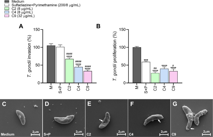
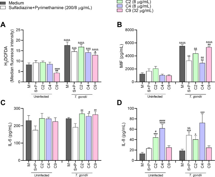
Congenital toxoplasmosis is a significant public health issue caused by the transplacental passage of Toxoplasma gondii to the embryo/fetus. The standard treatment involves a combination of sulfadiazine and pyrimethamine, drugs often associated with adverse effects and high toxicity. The current study aimed to investigate the potential of prenylated chalcones (C2, C4 and C9) in controlling T. gondii infection in human trophoblast cells (BeWo) and human placental explants. As results, non-cytotoxic doses of C2, C4 and C9 impaired parasite invasion and subsequent intracellular proliferation in BeWo cells. Scanning and transmission electron microscopies evidenced the direct effect of chalcones on tachyzoites, which presented irregular rough surface, membrane with hole-like structures, torsion and shape substantial changes after pretreatment. C4 and, especially C9, caused notable ultrastructural damages due to the formation of vacuole-like structures in the parasite cytoplasm and surrounding the parasitophorous vacuole. Additionally, chalcones modulated the cytokine profile by increasing IL-8 and downmodulating MIF and ROS levels in BeWo cells and downregulating TNF-α release in villous explants. These findings highlight C2, C4, and C9 as promising candidates for the development of alternative therapies to prevent congenital toxoplasmosis, as well as chalcones as a valuable scaffold for the design of new anti-T. gondii agents.

Keywords: Toxoplasma gondii; Chalcones; Flavonoid; Placenta; Treatment; Trophoblast.


Conflict of interest statement

Declarations. Competing interests: The authors declare no competing interests and no competing financial interests.

Figures

Fig. 1
Fig. 1
Cell viability and T. gondii intracellular proliferation assays. (AC): BeWo cells were treated with different concentrations of C2 (A), C4 (B) and C9 (C) (0.5–128 µg/mL) for cell viability analysis (MTT colorimetric assay). Cells were also treated only with culture medium (M), representing the negative control (100% of viability), and with DMSO 0.25%, representing the vehicle control (D). The absorbance was measured at 570 nm and cell viability was expressed in percentages (cell viability %). (D–F): β-galactosidase colorimetric assays were performed to quantify T. gondii intracellular proliferation in infected BeWo cells treated with non-cytotoxic concentrations of chalcones. Infected cells treated only with culture medium (M) represented the negative control (100% parasite proliferation), and treatment with a combination of 200 µg/mL of sulfadiazine and 8 µg/mL of pyrimethamine (S + P) represented the positive control. The absorbance was measured at 570 nm and T. gondii intracellular proliferation was expressed in percentages (% of T. gondii proliferation). (A–C): *Comparison between untreated and treated cells. (D–F): *Comparison between infected/treated and infected/untreated cells; #Comparison between infected/treated and S + P/infected cells. Data are presented as means ± SEM from three independent experiments performed in eight replicates. Significant differences (P < 0.05) were detected by the One-Way ANOVA with Dunnett’s multiple comparison post-test.
Fig. 2
Fig. 2
Reversibility assay. Infected BeWo cells were exposed to C2 (8 µg/mL), C4 (8 µg/mL), C9 (32 µg/mL), S + P (200 + 8 µg/mL) or only culture medium (M) for 24 h, followed or not by treatment removal for an additional 24 h. The β-galactosidase assay was performed and T. gondii intracellular proliferation was expressed in percentages (% of T. gondii proliferation). Treatment reversibility was deliberate by comparing the conditions of 24 h of treatment with the 24 h after treatment removal. The negative control group (M) was considered as 100% reversible. Data are expressed as means ± SEM from three experiments performed in eight replicates. (*) Comparisons between infected/untreated and infected/treated cells; (#) S + P and chalcone-treated cells; and (&) between infected/treated cells 24 h after treatment with the infected/treated cells 24 h after treatment removal. Significant differences were analyzed using (*;#) One-Way ANOVA with Dunnett’s multiple comparison post-test and (&) Unpaired Student´s t-test (two-tailed). Differences were considered as statistically significant when P < 0.05.
Fig. 3
Fig. 3
Ultrastructure analyses of T. gondii tachyzoites in the presence of chalcones. Transmission electron microscopy (TEM) micrographs showing T. gondii tachyzoites in BeWo cells treated for 24 h with: (A) only culture medium, (B) C2 (8 µg/mL), (C) C4 (8 µg/mL) and (D, E) C9 (32 µg/mL). Arrowhead: double membrane of the parasite. Black arrows: Vls (vacuole-like structures). Black asterisks: host cell mitochondria. Dg: dense granule. Nu: host cell nucleus. Rp: rhoptries. PNu, parasite nucleus; PV, parasitophorous vacuole. Scale bars (bottom left) 2 μm.
Fig. 4
Fig. 4
Chalcones-pretreated parasites adhesion in BeWo cells. T. gondii tachyzoites were pre-incubated for 1 h with C2 (8 µg/mL), C4 (8 µg/mL), C9 (32 µg/mL), S + P (200 + 8 µg/mL) or only culture medium (M) and then allowed to adhere in previously fixed BeWo cells for 3 h. Cells and parasites were fixed and labeled with DAPI (nuclei - blue), Phalloidin-Atto 565 (F-actin - red) and Alexa Fluor 488-conjugated anti-mouse IgG (tachyzoites – green). Images were captured using a confocal microscopy and approximately 20 fields were examined randomly to obtain: (A): the number of cells with adhered parasites. (B): the total number of adhered parasites per field analyzed. Significant differences (P < 0.05) were analyzed using Kruskal–Wallis test and Dunn’s multiple comparison post-test. Representative images highlighting the tachyzoites-host cell interaction of the different experimental conditions, as follow: (C) untreated parasites, (D) S + P-pretreated parasites, and (E, F, G) parasites pretreated with C2, C4 and C9, respectively. Data are expressed as means ± SEM from two independent experiments performed in four replicates. *Comparison between infected/untreated and infected/treated cells. #Comparison to S + P-treated cells. Scale bar: 40 μm. Tachyzoites were indicated by white arrowheads.
Fig. 5
Fig. 5
Chalcones-pretreated parasites. A pretreatment was carried out by incubating T. gondii tachyzoites with C2 (8 µg/mL), C4 (8 µg/mL), C9 (32 µg/mL), S + P (200 + 8 µg/mL) or only culture medium for 1 h. (A): β-galactosidase assay was performed 3 h later to obtain T. gondii invasion rate (% of T. gondii invasion). Significant differences (P < 0.05) were analyzed using one-way ANOVA and Dunnett´s multiple comparisons post-test. (B): β-galactosidase assay was performed 24 h later and T. gondii proliferation was expressed in percentages (% of T. gondii proliferation). Significant differences (P < 0.05) were analyzed using Kruskal–Wallis test and Dunn’s multiple comparison post-test. Untreated parasites (M) represent the negative control and were considered as 100% of invasion and proliferation, respectively. Data are expressed as means ± SEM from three experiments performed in eight replicates. *Comparison between untreated/infected and infected/treated cells. #Comparison to S + P-treated cells. Representative scanning electron microscopy (SEM) images showing: (C) untreated T. gondii tachyzoite, (D) tachyzoite pretreated with S + P (200 + 8 µg/ml), (E) C2 (8 µg/mL), (F) C4 (8 µg/mL) and (G) C9 (32 µg/mL). Scale bars (bottom right) 2 μm. Loss of tachyzoite surface integrity or hole-like structures was indicated by white arrows.
Fig. 6
Fig. 6
ROS and cytokine production in BeWo cells treated with chalcones. BeWo cells were infected or not with T. gondii tachyzoites, followed by a 24 h treatment with C2 (8 µg/mL), C4 (8 µg/mL), C9 (32 µg/mL), S + P (200 + 8 µg/mL) or only culture medium (M). (A): BeWo cells were incubated with the probe 2′,7′-dichlorodihydrofluorescein diacetate (H2DCF-DA) and the ROS production was measured by fluorescence using a spectrophotometer. Data were expressed as MFI. After treatments, the supernatants were collected for measurement of (B) MIF, (C) IL-6 and (D) IL-8 cytokines. Data are expressed as means ± SEM from four independent experiments performed in twelve replicates. *Comparison between untreated and treated-cells. #Comparison between S + P and chalcone-treated cells. &Comparison between infected/treated cells with the respective control in the absence of infection. (A, C): significant differences (P < 0.05) were analyzed using (*;#) One-Way ANOVA with Dunnett’s multiple comparison post-test and (&) Unpaired Student´s t-test (two-tailed). (B, D): significant differences were analyzed using (*;#) Kruskal–Wallis test and Dunn’s multiple comparison post-test and (&) Mann-Whitney test (two-tailed).
Fig. 7
Fig. 7
Tissue viability assays in human chorionic villous explants treated with chalcones. Villous explants were treated for 24 h with different concentrations of C2, C4 and C9 (0.5–128 µg/mL) for tissue viability analysis. In parallel, tissues were also treated with culture medium only (M), representing the negative control (100% of viability) and with DMSO 0.25% (D), representing the vehicle control. (A, B, C): MTT colorimetric assay was performed and expressed in percentages (viability % by MTT incorporation). (D, E, F): supernatants were collected and used to measure LDH (U/L) levels (LDH assay). Data are means ± SEM from eight experiments performed in eight replicates. *Comparison between untreated and treated villi. Significant differences (P < 0.05) were determined using One-Way ANOVA and Dunnett’s multiple comparisons post-test. Representative photomicrographs of hematoxylin–eosin (HE) stained histological sections of villi incubated with culture medium (G), C2 (H), C4 (I) and C9 (J) (all 64 µg/mL). Syncytiotrophoblast cells are indicated by black arrows and mesenchyme by letter M. Scale bar: 60 μm.
Fig. 8
Fig. 8
T. gondii intracellular proliferation and cytokine production in human villous explants treated with chalcones. (A) Villous explants were infected with T. gondii tachyzoites and treated with C2, C4, C9 (32 and 64 µg/mL), S + P (150 + 200 µg/mL, respectively) or culture medium only for 24 h. The percentages of T. gondii intracellular proliferation were measured using the β-galactosidase assay and compared with infected/untreated explants (M), considered as 100% of parasite proliferation, and positive control (S + P). B, C: After treatments, supernatants were collected for measurement of (B) MIF and (C) TNF-α. Cytokine levels were expressed in pg/mg of tissue. Data are expressed as means ± SEM of three independent experiments performed in eight replicates. *Comparison between untreated and treated villi. #Comparison to S + P-treated cells. &Comparison between uninfected/treated and infected/treated explants. Significant differences were analyzed using (*;#) One-Way ANOVA with Dunnett’s multiple comparison post-test and (&) Unpaired Student´s t-test (two-tailed). Differences were considered as statistically significant when P < 0.05.
Fig. 9
Fig. 9
Synthesis of compounds C2, C4 (A) and C9 (B).

Similar articles

References

    1. Dubey, J. P., Lago, E. G., Gennari, S. M., Su, C. & Jones, J. L. Toxoplasmosis in humans and animals in brazil: High prevalence, high burden of disease, and epidemiology. Parasitology139, 1375–1424. 10.1017/s0031182012000765 (2012). - PubMed
    1. Montoya, J. G., Liesenfeld, O. & Toxoplasmosis Lancet 363, 1965– (1976). 10.1016/s0140-6736(04)16412-x (2004). - PubMed
    1. Ahmed, M., Sood, A. & Gupta, J. Toxoplasmosis in pregnancy. Eur. J. Obstet. Gynecol. Reprod. Biol.255, 44–50. 10.1016/j.ejogrb.2020.10.003 (2020). - PubMed
    1. Kodjikian, L. et al. Vertical transmission of toxoplasmosis from a chronically infected immunocompetent woman. Pediatr. Infect. Dis. J.23, 272–274. 10.1097/01.inf.0000115949.12206.69 (2004). - PubMed
    1. Li, X. L., Wei, H. X., Zhang, H., Peng, H. J. & Lindsay, D. S. A Meta analysis on risks of adverse pregnancy outcomes in Toxoplasma gondii infection. PLoS One. 9, e97775. 10.1371/journal.pone.0097775 (2014). - PMC - PubMed