. 2025 Aug;644(8076):490-496.
doi: 10.1038/s41586-025-09229-y. Epub 2025 Jul 2.

Electron flow in hydrogenotrophic methanogens under nickel limitation

Affiliations

Electron flow in hydrogenotrophic methanogens under nickel limitation

Shunsuke Nomura et al. Nature. 2025 Aug.

Abstract

Methanogenic archaea are the main producers of the potent greenhouse gas methane1,2. In the methanogenic pathway from CO2 and H2 studied under laboratory conditions, low-potential electrons for CO2 reduction are generated by a flavin-based electron-bifurcation reaction catalysed by heterodisulfide reductase (Hdr) complexed with the associated [NiFe]-hydrogenase (Mvh)3-5. F420-reducing [NiFe]-hydrogenase (Frh) provides electrons to the methanogenic pathway through the electron carrier F420 (ref. 6). Here we report that under strictly nickel-limited conditions, in which the nickel concentration is similar to those often observed in natural habitats7-11, the production of both [NiFe]-hydrogenases in Methanothermobacter marburgensis is strongly downregulated. The Frh reaction is substituted by a coupled reaction with [Fe]-hydrogenase (Hmd), and the role of Mvh is taken over by F420-dependent electron-donating proteins (Elp). Thus, Hmd provides all electrons for the reducing metabolism under these nickel-limited conditions. Biochemical and structural characterization of Elp-Hdr complexes confirms the electronic interaction between Elp and Hdr. The conservation of the genes encoding Elp and Hmd in CO2-reducing hydrogenotrophic methanogens suggests that the Hmd system is an alternative pathway for electron flow in CO2-reducing hydrogenotrophic methanogens under nickel-limited conditions.


Conflict of interest statement

Competing interests: The authors declare no competing interests.

Figures

Fig. 1
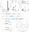
Fig. 1. Proteomic analysis of M.marburgensis cells under limiting conditions.
a, The table at the top shows the reduction rate (%) of the Ni2+ and Fe2+ concentrations, and the flow rate of H2/CO2 gas in the limitation culture (Conc.), and the specific growth rate (μ (h−1)) of the cultures used for proteomic analysis. Ni2+ and Fe2+ in the standard condition are 5 μM and 50 μM, respectively. The total intensity of the mass spectrometry-based proteomic analysis (Total) and the intensity of the individual proteins of three distinct samples are calculated as a percentage of the values obtained under the standard culture condition and the relative intensities are shown by a heat map. Proteins related to the CO2-reducing hydrogenotrophic methanogenic pathway are shown. Ftr, formyltransferase; Mch, cyclohydrolase; Mer, methylene-H4MPT reductase; Transporter, MTBMA_c10530; ABC transporter, MTBMA_c10830; Feo, ferrous iron transport protein. *15–50% or 200–400%; **5–15% or 400–1,000%; ***<5% or >1,000%. bh, The proteomic intensity of MvhA, MvhB, MvhD, MvhG (b), ElpA, ElpB, ElpC, ElpX (c), HdrA, HdrB, HdrC (d), FrhA, FrhB, FrhG (e), Mtd (f), Hmd (g) and total (h) from samples from the cells cultivated under various nickel conditions (5,000 nM, 250 nM, 125 nM and 50 nM). Means of the proteome intensity of three distinct samples are shown and error bars indicate standard error (s.e.). Source data
Fig. 2
Fig. 2. Enzymological analysis of the Hdr complex from M. marburgensis cells and purification of the Elp–Hdr complex.
ac, Activity of the enzymes in the cell extract. Means of three measurements of distinct samples obtained from the same cell extract (n = 1) are shown and error bars indicate s.e. a, The enzyme activity under nickel-sufficient and nickel-limiting conditions. b, H2-dependent Hdr activity is shown by production of thiols (CoM-SH and CoB-SH) from CoM-S-S-CoB using cell extract. The cell extracts from the nickel-sufficient culture (5 µM Ni2+; filled label) and nickel-limiting culture (50 nM Ni2+; open label) were tested under either H2 or N2. c, H2-dependent Hdr activity of the washed cell extracts from the nickel-limiting cultured cells was tested. To confirm the activity, addition of the Hmd-specific inhibitor toluenesulfonylmethyl isocyanide (TosMIC) and the conditions containing only one substrate (without F420 or without methenyl-H4MPT+) or lacking both substrates (no addition of substrates) were tested. d, Gene clusters encoding the proteins associated with Hdr. bp, base pairs. e, FBEB reaction catalysed by the Mvh–Hdr complex from M.marburgensis under nickel-sufficient conditions and the Elp–Hdr complex from M.marburgensis under nickel-limiting conditions. f, Characterization of the fractions eluted from size-exclusion chromatography of the Hdr complex from M.marburgensis cells from nickel-limiting continuous culture (50 nM Ni2+). Hmd+Mtd-coupled-reaction-mediated F420-dependent Hdr (F420 Hdr) activity and benzyl-viologen-dependent Hdr (BV Hdr) activity are shown. Absorbance at 280 nm (arbitrary units) is shown as a dashed line. Source data
Fig. 3
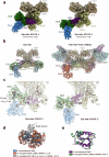
Fig. 3. Structure and reaction of the Elp–Hdr complex from M. marburgensis.
a,b, In the Elp–Hdr complex (a), the ElpABC subunits interact with the Hdr dimer in a manner similar to that observed in the Mvh–Hdr dimer composed of MvhAG, MvhD and HdrABC from M. thermolithotrophicus (b; Protein Data Bank (PDB) accession code 5ODH). F420H2 is oxidized at the ElpB FAD-binding site, and electrons are transferred through the cubane [4Fe–4S] clusters of ElpB (EB(1, 2, 4)) and of the N terminus of HdrA ((HA(1,2)) to reach the [2Fe–2S] cluster of ElpC (EC), from which they are presumably transferred to the bifurcating HdrA′ FAD′ by an unknown mechanism (Extended Data Fig. 5c).
Fig. 4
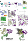
Fig. 4. Transition of the hydrogenase and electron-donating system in methanogenesis from CO2 and H2 under strictly nickel-limiting conditions.
a,b, Schematic views of the methanogenic pathway under nickel-sufficient (a) and nickel-limiting (b) conditions. Two [NiFe]-hydrogenases, Frh and Mvh, are strongly downregulated under the strictly nickel-limiting conditions. Their functions are substituted by the coupled enzyme system of [Fe]-hydrogenase (Hmd) with Mtd, and Elp complexed with Hdr. The drawing of Hmd and Mtd in b indicates a coupled reaction rather than a complex formation. Figure adapted from ref.  under a Creative Commons licence CC BY 4.0.
Extended Data Fig. 1
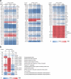
Extended Data Fig. 1. Proteomic analysis of Methanothermobacter marburgensis cells under limiting conditions.
a. Change of the expression under 250 nM, 125 nM, and 50 nM Ni2+ concentrations. The control culture contained 5 µM Ni2+. Means of the proteome intensity of distinct samples (n = 3) are shown and error bars indicate standard error (SE). Membrane-associated [NiFe]-hydrogenases (Eha and Ehb). CO dehydrogenase (Cdh), 30S ribosomal protein (Rps), 50S ribosomal protein (Rpl). For other abbreviations, see Fig. 1 of the main text. The up-regulation of production of “Transporter” and “ABC transporter” suggests that these proteins could be transporters involved in Ni transport. b. In this figure, proteins that are not involved in the CO2-reducing hydrogenotrophic methanogenic pathway but exhibited changes in protein intensity are shown. The concentration (Conc) of Ni2+ and Fe2+, the total intensity of the mass spectrometry-based proteomic analysis (Intensity) and the proteome intensity of the individual proteins are shown as a percentage of the values obtained under the standard culture condition (Control), in which the concentration of Ni2+ and Fe2+ in the control are 5 μM and 50 μM, respectively. Means of three distinct samples are shown. The specific growth rate (μ) was calculated from the dilution rate of the continuous flow rate of the cultures used for the proteomic analysis. The product of the genes up-regulated under the Ni2+ and Fe2+-limited condition might be involved in transport of the respective metal ion. Source data
Extended Data Fig. 2
Extended Data Fig. 2. SDS-PAGE of the size exclusion chromatography (Superose 6 Increase) fractions.
(Left) Fractions 1–13 every 0.5 ml starting at 11.25 ml, shown in Fig. 2f, were analyzed by SDS-PAGE using 4 -16 % gradient polyacrylamide gel from Bio-Rad Laboratories. The eluted fraction was concentrated 10-fold through a 3-kDa ultrafilter and 10 μl were subjected to denaturation in SDS and loaded onto the gel. (M) Marker proteins with the molecular mass. (Right) The SDS-PAGE lane of fraction 4 containing the 1-MDa complex is shown to indicate the deduced position of the subunits of Elp, Hdr, and Fmd (Fwd). Four experiments were repeated independently with similar results (for the uncropped data for the gel, see Supplementary Fig. 7).
Extended Data Fig. 3
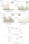
Extended Data Fig. 3. Proteomic analysis of the size-exclusion chromatography step of fractionation of the Elp-Hdr-Fmd complex.
a-d. Mass spectrometry-based proteomic analysis of the size-exclusion chromatography step of fractionation of the Elp-Hdr-Fmd complex shown in panel Fig. 2f. HdrABC (a), ElpABC (b), FwdA, FmdB, and FmdC (c), and MvhAGDB (d). The total number of identified peptide spectra matches for the protein (PSM) (n = 1) is shown as the intensity of the proteins in the ordinate. e. Purification from the nickel-limiting cells (50 nM Ni2+), from disruption of the cells through the three chromatographic steps, was completed within 1 day. f, Re-chromatography of the 1-MDa fraction from panel e after 40 h storage on ice. Although stability testing of the Elp-Hdr-Fmd complex of the purified 1-MDa complex was only conducted once, the elution profiles from the multiple purification processes, which differed in terms of the size of the 1-MDa peak, support that the complex is unstable. Source data
Extended Data Fig. 4
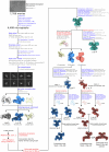
Extended Data Fig. 4. Image processing workflow for the analysis of the Elp-Hdr dataset.
State 1 and State 2 correspond to the two different conformational states of the mobile arm composed of ElpA, B and C, and the N- and C-terminal regions of HdrA. Resolutions were estimated using the gold-standard FSC at 0.143. Steps carried out in CryoSPARC are shown in blue, whereas steps carried out in Relion 4.0 are shown in red. Abbreviations used: External (ext.), pixel (px), per (p.), resolution (res.), particles (ptcls), including (incl.), extension (ext.), soft-edge (soft), classification (class.), iterations (it.), excluding (excl.), spherical aberration (spher. abb.), anisotropic magnification (anis. mag.) and B-factor (Bfac.).
Extended Data Fig. 5
Extended Data Fig. 5. The Elp-Hdr complex of M. marburgensis is structurally similar to the Fdh-Hdr region of the Fdh-Hdr-Fmd complex of M. hungatei.
a, Composite maps of the two different conformational states of the Elp-Hdr complex, which are very similar to the states 1 and 2 described for Fdh-Hdr-Fmd in M. hungatei. b, The structure of the Elp-Hdr complex (left) is highly similar to the Fdh-Hdr region of the Fdh-Hdr-Fmd complex (right) of M. hungatei (PDB:7BKC). c, Atomic models of the conformational states 1 (left) and 2 (right) of the Elp-Hdr mobile arm composed of subunits ElpA (blue), B (green) and C (purple), and the N- and C-terminal domains of HdrA (khaki). Following the proposed model of electron transfer in Fdh-Hdr-Fmd, in Elp-Hdr State 1 the [2Fe-2S] cluster of ElpC (EC) moves closer to the bifurcating FAD’ of HdrA’ (25 Å distance) to transfer two electrons through an unknown mechanism. The complex transitions to State 2 through the rotation of the mobile arm, by which the [4Fe-4S] cluster of the HdrA C terminus (HA3) moves closer to the reduced FAD’ (20 Å distance). The high-potential electron is transferred from the hydroquinone state of FAD’ to CoM-S-S-CoB via HA4’, whereas the low-potential electron from the flavosemiquinone state is transferred to HA3, the ‘shuttle cluster’. A transition back to conformational state 1 brings HA3 near enough to HA5’ for efficient electron transfer. d, ElpA (blue) is highly similar to FdhA (PDB:7BKC, orange), but lacks the molybdopterin-binding (MopB) domain and is inactive for the Fdh reaction. The AlphaFold2 model of M. hungatei FdhA (AF-Q2FRK1-F1) is also displayed (grey) to show the MopB domain, which was not deposited for M. hungatei FdhA (orange) due to low map resolution; however, unlike for the Elp complex, clear density was observed for the MopB domain of M. hungatei FdhA. e, ElpC is highly similar to M. hungatei MvhD and shows the [2Fe-2S] cluster (EC) for electron transfer to the bifurcating FAD of HdrA.
Extended Data Fig. 6
Extended Data Fig. 6. Comparison of the structure of the HdrABC dimer and ElpB from M. marburgensis with their homologs.
a, The structure of the core (HdrABC)2 region in the Elp-Hdr complex is highly similar to the (HdrABC)2 of M. hungatei (Fdh-Hdr-Fmd complex, PDB:7BKC, root-mean-square deviation (RMSD) of 0.952 Å between 516 amino acids) and the (HdrABC)2 of M. thermolithotrophicus (Mvh-Hdr complex, PDB:5ODH, RMSD of 0.659 Å between 390 amino acids). b, The ElpB subunit of the Elp-Hdr complex is homologous to the FdhB subunit of the FdhAB-MvhD arm of M. hungatei (PDB:7BKC, RMSD 0.783 Å between 328 amino acids).
Extended Data Fig. 7
Extended Data Fig. 7. MvhB interacts with the inserted ferredoxin-like domain of HdrA.
a, 3D classification without alignment masking the AF3-predicted MvhB subunit revealed around 12.1 % of the C2-expanded particles show an additional density bound to the inserted ferredoxin-like domain of HdrA. The map could be refined to 2.58 Å. Processing steps performed in CryoSPARC are shown in blue, whereas steps performed in RELION are shown in red. b, AF3-predicted structure of M. marburgensis MvhB. The structure is colored according to the per-atom predicted local distance difference test (pLDDT) scores. Predicted alignment error (PAE) scores are also shown. c, AF3-predicted MvhB (N-terminal residues 1-57, 67-124) can be fitted into the refined map. The [4Fe-4S] clusters HA6 and MB3 connect HdrA and MvhB electronically (9.8 Å distance). d, The AF3-predicted N-terminal region of MvhB displays a mean Q-score of 0.61 when fitted into the map. e, Detail of the densities and fitted [4Fe-4S] clusters (MB1 to MB4) present in the resolved N-terminal region of MvhB. Coordinating cysteines are shown as sticks. f, The AF3 structure of HdrA-MvhB-FmdF predicts that MvhB mediates the interaction between Elp-Hdr and Fmd. g, The AF3-predicted HdrA (khaki) and MvhB (purple) subunits can be correctly fitted into the HdrA-MvhB map. Other abbreviations used: binarization (bin.), external (ext.), pixel (px), low-pass filtered (lp.), blush regularization (blush reg.), particles (ptcls), extension (ext.), soft-edge (soft), predicted template modeling AF3 score (pTM), interface predicted template modeling AF3 score (ipTM).
Extended Data Fig. 8
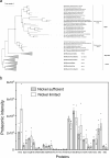
Extended Data Fig. 8. Phylogenetic tree of ElpA and its homologs and the proteomic intensity of samples from cells of Methanothermococcus thermolithotrophicus.
a. Phylogenetic tree of ElpA and its homologs, ElpA-like and FdhA. ElpA, ElpA-like, and FdhA can be separated based on phylogeny and the sequence length (AA, amino-acid sequence length). AlphaFold prediction indicated that the tertiary structure of ElpA-like proteins of Class II methanogens (Methanosarcinales and Methanomicrobiales) shows higher similarity to that of FdhA. Exceptionally, the tertiary structure of ElpA-like from Methanocorpusculum species is structurally almost identical to ElpA from Class I methanogens (Methanobacteriales and Methanococcales) (see also Supplementary Table 1). The maximum-likelihood tree is based on a MUSCLE alignment and was generated using IQ-TREE with LG + I + G4 model of evolution. Node support was tested with nonparametric bootstrap analysis (filled circles indicate support 70% or higher; 100 replicates). The scale bar indicates the number of substitutions per site. The tree was rooted with various bacterial FdhA. b. The proteomic intensity from M. thermolithotrophicus (DSM 2095) cultivated under nickel-sufficient (5 μM Ni2+) and nickel-limited (50 nM) conditions. Means of the proteome intensity (iBAQ) of the three distinct samples are shown and error bars indicate standard error (SE) (n = 3). Hmd, WP_018153721.1; ElpA, WP_018153231.1; ElpB, WP_018153230.1; MvhA, WP_018154262.1; MvhG, WP_018154261.1; MvhD, WP_018154260.1; MvhB, WP_018154263.1; FrhA, WP_018154259.1; FrhG, WP_018154257.1; FrhB, WP_018153424.1; McrA, WP_018153522.1; McrB, WP_018153526.1; McrG, WP_018153523.1; HdrA, WP_018154264.1; HdrB, WP_018154154.1; HdrC, WP_018154153.1; Mer, WP_026182932.1; Mtd, WP_018154202.1. Proteome intensity of the subunits of the isoenzymes of MvhAGDB encoded in the VhuAGDB gene cluster (WP_018154225.1, WP_018154224.1, WP_245547903.1, and WP_018154226.1) was very low. Isoenzymes of HdrBC (WP_018154154.1 and WP_018154031.1) were not detected. Proteome intensity of McrABG (WP_018154763.1, WP_018154760.1, and WP_018154762.1) was much lower than the counterpart. Therefore, proteome intensity of these isoenzymes is not shown in this figure. Source data

Similar articles

References

    1. Conrad, R. The global methane cycle: recent advances in understanding the microbial processes involved. Environ. Microbiol. Rep.1, 285–292 (2009). - PubMed
    1. Thauer, R. K., Kaster, A. K., Seedorf, H., Buckel, W. & Hedderich, R. Methanogenic archaea: ecologically relevant differences in energy conservation. Nat. Rev. Microbiol.6, 579–591 (2008). - PubMed
    1. Setzke, E., Hedderich, R., Heiden, S. & Thauer, R. K. H2: heterodisulfide oxidoreductase complex from Methanobacterium thermoautotrophicum: composition and properties. Eur. J. Biochem.220, 139–148 (1994). - PubMed
    1. Kaster, A. K., Moll, J., Parey, K. & Thauer, R. K. Coupling of ferredoxin and heterodisulfide reduction via electron bifurcation in hydrogenotrophic methanogenic archaea. Proc. Natl Acad. Sci. USA108, 2981–2986 (2011). - PMC - PubMed
    1. Wagner, T., Koch, J., Ermler, U. & Shima, S. Methanogenic heterodisulfide reductase (HdrABC-MvhAGD) uses two noncubane [4Fe-4S] clusters for reduction. Science357, 699–702 (2017). - PubMed

LinkOut - more resources