. 2025 Jul;29(13):e70686.
doi: 10.1111/jcmm.70686.

Robust Increase in IQCK Protein Expression in Mouse Models of Alzheimer's Disease and iPSC-Derived Neurons

Affiliations

Robust Increase in IQCK Protein Expression in Mouse Models of Alzheimer's Disease and iPSC-Derived Neurons

Juliet Akkaoui et al. J Cell Mol Med. 2025 Jul.

Abstract

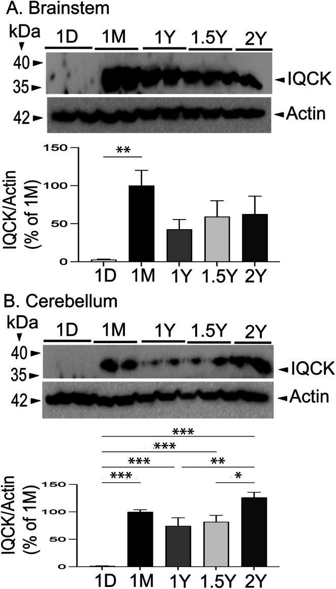
Emerging studies indicate that the IQ-motif-containing protein K (IQCK) is a novel risk factor for Alzheimer's disease (AD), an age-associated disease. The expression patterns of IQCK in healthy and AD brains, within the context of age and sex are largely unknown. Therefore, we compared the age-dependent expression patterns of IQCK in males and females of wild-type (WT) mice with AD-like 3xTg and APΔE9 mice. Additionally, we measured IQCK protein expression in AD-derived human iPSC neurons. In WT mice, we found no IQCK expression at day 1 (1D) in the cortex (CX), hippocampus (HP), brainstem (BS) and cerebellum (CB). Overall, IQCK protein expression in different brain regions was first detected in 1-month-old wild-type (WT) mice, reaching its maximum in 1-year-old mice (1Y), and then gradually decreased in 2-year-old mice. In the APΔE9 mice, IQCK protein levels significantly increased by 1246% in the CX, 682% in the HP and 169% in the BS relative to WT controls. In the 3xTg mice, only HP showed an increase of IQCK protein by 277%. In addition, we also detected elevated tendencies in BS and CB regions but not in the CX. Finally, IQCK expression was also significantly increased by 68% in the AD-derived iPSC neurons relative to the NC-derived iPSC neurons. Thus, increased IQCK protein levels in the brain of AD-like 3xTg and APΔE9 mouse models suggest a possible role in AD pathogenesis, a finding that requires further clarification.

Keywords: 3xTg mice; APΔE9 mice; Aging; Alzheimer's disease; Brain regions; Dendrites; IQCK; iPSC neurons.


Conflict of interest statement

The authors declare no conflicts of interest.

Figures

FIGURE 1
FIGURE 1
Age‐dependent variations in the expression of IQCK protein in the cortex (CX) and hippocampus (HP) of wild‐type (WT) mice at 1 day (1D), 1 month (1M), 1 year (1Y), 1.5 years (1.5Y) and 2 years (2Y). In the CX, IQCK protein expression is absent at 1D, increases to maximum levels at 1Y, and then gradually reduces by 2Y. In the HP, IQCK protein levels reached a maximum at 1 year and then gradually decreased at other ages. Data were statistically analyzed by Analysis of Variance (ANOVA) followed by the Tukey–Kramer Multiple Comparisons Test. *, p < 0.05, **, p < 0.01 and ***, p < 0.001, Data are mean ± SEM, n = 3 per group.
FIGURE 2
FIGURE 2
Age‐dependent variations in the expression of IQCK protein in the brainstem (BS) and cerebellum (CB) of wild‐type (WT) mice at 1 day (1D), 1 month (1M), 1 year (1Y), 1.5 years (1.5Y) and 2 years (2Y). In the BS, IQCK protein expression is absent at 1D, increases to maximum levels at 1M, and then is expressed at lower levels at other ages. In the CB, no IQCK protein expression was observed at 1D, followed by comparable expression at 1M, 1Y and 1.5Y, with the highest expression at 2Y. Data were statistically analyzed by Analysis of Variance (ANOVA) followed by the Tukey–Kramer Multiple Comparisons Test. *, p < 0.05, **, p < 0.01 and ***, p < 0.001. Data are mean ± SEM, n = 3 per group.
FIGURE 3
FIGURE 3
IQCK protein is significantly increased in different regions of the brain in the APΔE9 mouse model of AD relative to wild‐type (WT; non‐transgenic) controls, as detected by western blot assay. IQCK protein levels were normalized to Actin levels, used as loading controls, and quantified using ImageJ, and then compared among APΔE9 and WT control mice. APP expression is shown to validate the APΔE9 mice relative to WT controls. The APΔE9 cortex (CX) showed 1246% increased IQCK levels compared to WT mice. The hippocampus (HP) showed a 682% increase while the brainstem (BS) showed a 169% increase in the APΔE9 mice relative to WT controls. Data were statistically analyzed by paired t‐test. *, p < 0.05, **, p < 0.01 and ***, p < 0.001. Data are mean ± SEM, n = 3 per group.
FIGURE 4
FIGURE 4
IQCK protein is significantly increased in the hippocampus brain region of 3xTg AD‐like mice relative to the wild‐type (WT; non‐transgenic) controls, as detected by western blot assay. IQCK protein levels were normalized to Actin levels used as loading controls, quantified by ImageJ, and compared among 3xTg and WT control mice. APP expression is also shown to validate the 3xTg mice relative to WT controls. Relative to WT, the 3xTg hippocampus (HP) showed a 277% increase in IQCK levels. The other brain regions showed an increased trend but were not significant. Data were statistically analyzed by paired t‐test. *, p < 0.05. Data are mean ± SEM, n = 3 per group.
FIGURE 5
FIGURE 5
IQCK protein is expressed in progressively networking iPSC‐derived NeuN‐confirmed neurons and MAP2 and spinophilin confirmed dendritic spines. iPSC cells were grown and differentiated into neurons for 16 days in vitro (16DIV) and then immunocytochemically stained for IQCK, along with NeuN or Spinophilin, as markers for neurons or dendritic spines, respectively. (A) The gradual formation of a complex network of neurons is shown on DIV2, DIV8 and DIV14. (B) Staining with MAP2 antibody shows a complex network of dendrites. (C) IQCK protein is expressed in mature NeuN+ neurons. (D) IQCK protein is expressed in the dendritic spines, as supported by the merged images of IQCK and spinophilin.
FIGURE 6
FIGURE 6
IQCK protein expression is increased in iPSC‐derived neurons from AD patients relative to NC‐derived neurons. (A) iPSC neurons express APP and phosphor‐tau proteins. Confirmation of APP expression with two antibodies, 63D and CT15, and expression of phospho‐tau with AT100 antibody in iPSC‐derived neurons. (B) IQCK expression is increased in AD‐derived iPSC neurons relative to iPSC neurons derived from healthy controls. Quantification of IQCK immunofluorescence showed increased levels in AD‐derived iPSC neurons when compared to NC‐derived neurons. Data were statistically analyzed by paired t‐test. **, p < 0.01, Data are mean ± SEM, n = 4 per group.

Similar articles

References

    1. Moss M. B., Killiany R. J., Lai Z. C., Rosene D. L., and Herndon J. G., “Recognition Memory Span in Rhesus Monkeys of Advanced Age,” Neurobiology of Aging 18, no. 1 (1997): 13–19. - PubMed
    1. Herndon J. G., Moss M. B., Rosene D. L., and Killiany R. J., “Patterns of Cognitive Decline in Aged Rhesus Monkeys,” Behavioural Brain Research 87, no. 1 (1997): 25–34. - PubMed
    1. Goldberg A. L., “Protein Degradation and Protection Against Misfolded or Damaged Proteins,” Nature 426, no. 6968 (2003): 895–899. - PubMed
    1. Ohsumi Y., “Protein Turnover,” IUBMB Life 58, no. 5–6 (2006): 363–369. - PubMed
    1. Vilchez D., Saez I., and Dillin A., “The Role of Protein Clearance Mechanisms in Organismal Ageing and Age‐Related Diseases,” Nature Communications 5 (2014): 5659. - PubMed