

Emphysema: an ignored radiologic sign of diffuse alveolar hemorrhage
- PMID: 40604497
- PMCID: PMC12218831
- DOI: 10.1186/s12887-025-05850-y
Emphysema: an ignored radiologic sign of diffuse alveolar hemorrhage
Abstract
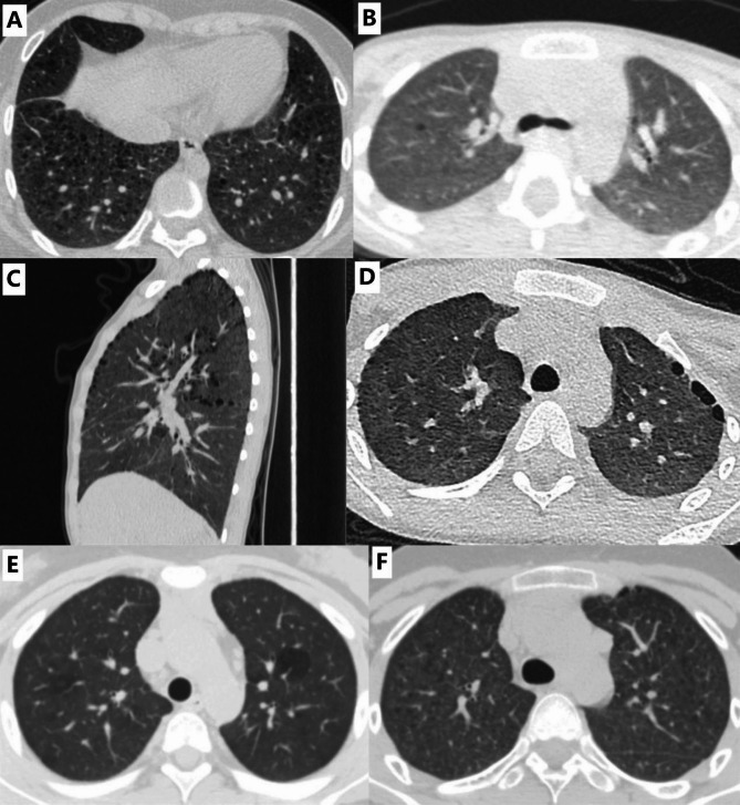
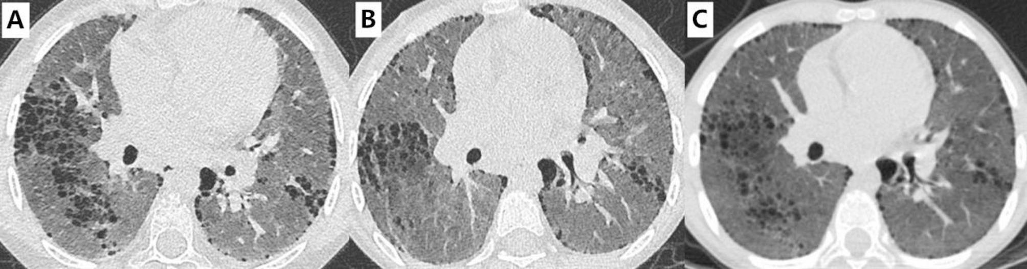
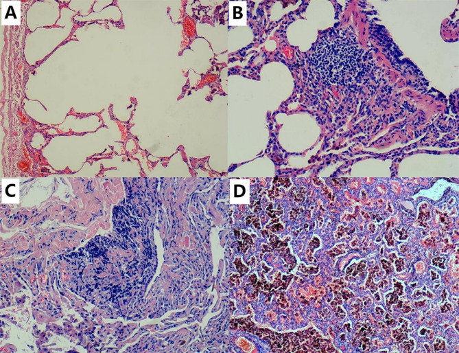
Background: Diffuse alveolar hemorrhage (DAH) is characterized by the presence of diffuse ground glass opacities and/or consolidation on chest CT. Emphysema has been reported occasionally. Nevertheless, the pathogenesis, incidence, imaging subtypes, and clinical impact of emphysema in patients with DAH are still unclear.
Methods: This was a retrospective clinical study. Children with DAH who were admitted to our hospital between January 2013 and December 2019 were included. All chest CT images of the patients were analyzed by two board-certified thoracic radiologists. The clinical features of the patients with diffuse emphysema were further analyzed. A matched case-control study was performed to explore the risk factors for developing diffuse emphysema.
Results: Ninety-four patients were included. Chest CT scans revealed emphysema in 28 patients (29.8%). Paraseptal emphysema (n = 24) was the most common imaging subtype. The median interval between the onset and development of emphysema was 11.0 months (n = 18, range: 2-113 months). Nine cases developed diffuse emphysema, in which six cases had persistent dyspnea and exercise intolerance, nine cases had persistent ground-glass opacities and 8 cases had a delay in initial treatment of 12 or more months. Compared to those in the matched control group, the interval between the onset and start of regular therapy with glucocorticoids was significantly longer in the cases with diffuse emphysema (median interval 13.5 vs. 1.5 months, P < 0.01).
Conclusions: In cases with DAH, emphysema is a relatively common but ignored radiologic sign and diffuse emphysema seems to be a sign of irreversible lung function impairment. A delay in initial treatment is a risk factor for developing diffuse emphysema.
Keywords: Chest CT; Delay of initial treatment; Diffuse alveolar hemorrhage; Emphysema.
© 2025. The Author(s).
Conflict of interest statement
Declarations. Ethical approval and consent to participate: The present study was reviewed and approved by the Ethics Committee of the First Affiliated Hospital of Guangxi Medical University (2023-E047-01). Informed consent was obtained from all the subjects and/or their legal guardian(s) in the case of minors (younger than 16 years of age). All the methods and procedures carried out in this study were in accordance with relevant guidelines and regulations. Consent for publication: Not applicable. Competing interests: The authors declare no competing interests.
Figures
Similar articles
-
Adenoidectomy for otitis media with effusion (OME) in children.Cochrane Database Syst Rev. 2023 Oct 23;10(10):CD015252. doi: 10.1002/14651858.CD015252.pub2. Cochrane Database Syst Rev. 2023. PMID: 37870083 Free PMC article.
-
Signs and symptoms to determine if a patient presenting in primary care or hospital outpatient settings has COVID-19.Cochrane Database Syst Rev. 2022 May 20;5(5):CD013665. doi: 10.1002/14651858.CD013665.pub3. Cochrane Database Syst Rev. 2022. PMID: 35593186 Free PMC article.
-
Lung volume reduction surgery for diffuse emphysema.Cochrane Database Syst Rev. 2016 Oct 14;10(10):CD001001. doi: 10.1002/14651858.CD001001.pub3. Cochrane Database Syst Rev. 2016. PMID: 27739074 Free PMC article.
-
Thoracic imaging tests for the diagnosis of COVID-19.Cochrane Database Syst Rev. 2022 May 16;5(5):CD013639. doi: 10.1002/14651858.CD013639.pub5. Cochrane Database Syst Rev. 2022. PMID: 35575286 Free PMC article.
-
Sertindole for schizophrenia.Cochrane Database Syst Rev. 2005 Jul 20;2005(3):CD001715. doi: 10.1002/14651858.CD001715.pub2. Cochrane Database Syst Rev. 2005. PMID: 16034864 Free PMC article.
References
-
- Newsome BR, Morales JE. Diffuse alveolar hemorrhage. South Med J. 2011;104:269–74. - PubMed
-
- Lara AR, Schwarz MI. Diffuse alveolar hemorrhage. Chest. 2010;137:1164–71. - PubMed
-
- Scapa JV, Fishbein GA, Wallace WD, Fishbein MC. Diffuse alveolar hemorrhage and pulmonary vasculitides: histopathologic findings. Semin Respir Crit Care Med. 2018;39(4):425–33. - PubMed
-
- Spira D, Wirths S, Skowronski F, Pintoffl J, Kaufmann S, Brodoefel H, et al. Diffuse alveolar hemorrhage in patients with hematological malignancies: HRCT patterns of pulmonary involvement and disease course. Clin Imaging. 2013;37(4):680–6. - PubMed
MeSH terms
Grants and funding
LinkOut - more resources
Full Text Sources
Medical
