. 2025 Jul 2;24(1):186.
doi: 10.1186/s12943-025-02391-x.

STK3 is a transcriptional target of YAP1 and a hub component in the crosstalk between Hippo and Wnt signaling pathways during gastric carcinogenesis

Affiliations

STK3 is a transcriptional target of YAP1 and a hub component in the crosstalk between Hippo and Wnt signaling pathways during gastric carcinogenesis

Fuda Xie et al. Mol Cancer. .

Abstract

Background: Serine/threonine kinase 3 (STK3) is recognized as a key regulator in Hippo pathway and a tumor-suppressing gene in various cancer types. However, its non-canonical role has been gradually revealed in cancer development.

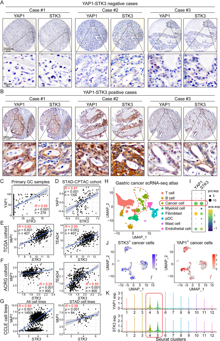
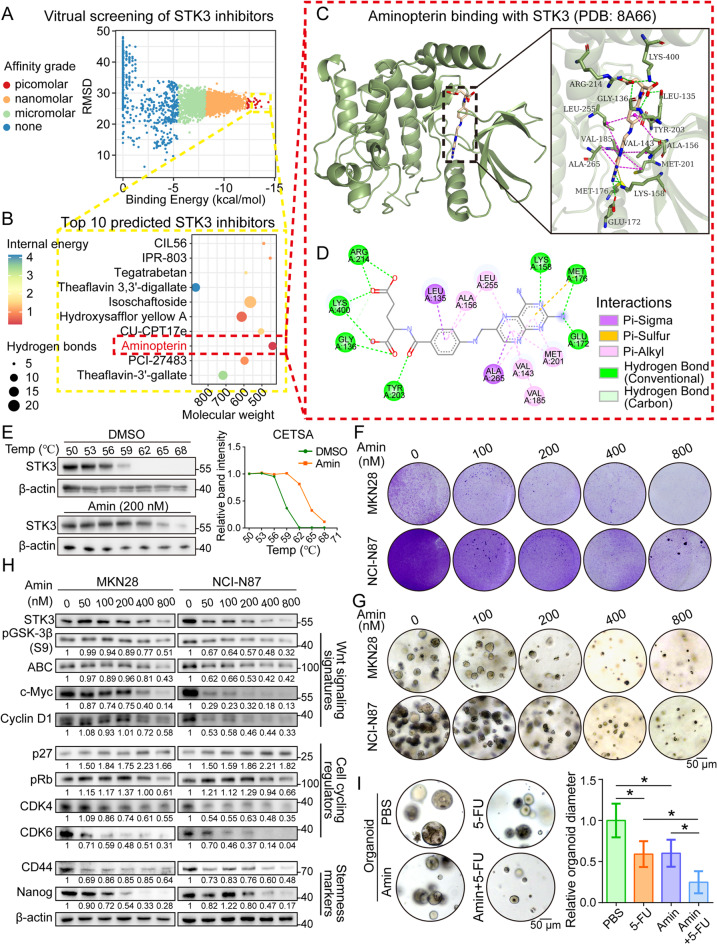
Methods: Our objective is to elucidate the upregulation pattern and molecular mechanisms of STK3 in advancing gastric cancer (GC) progression. The regulation of YAP1 on STK3 was assessed through a combination of bulk and single-cell RNA-sequencing, Western blot, ChIP-qPCR, gene knockout mouse models, and functional rescue assays. The oncogenic roles of STK3 were confirmed through subcutaneous xenograft formation models and functional assays including spheroid formation and organoid growth. The phosphorylated target of STK3 was revealed by co-immunoprecipitation and in vitro kinase assays. STK3-targeted drugs were screened out by molecular docking and cellular thermal shift assay (CETSA).

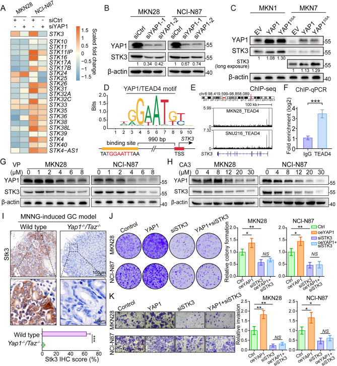
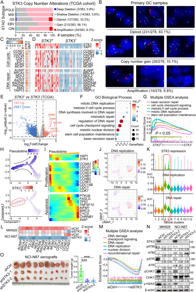
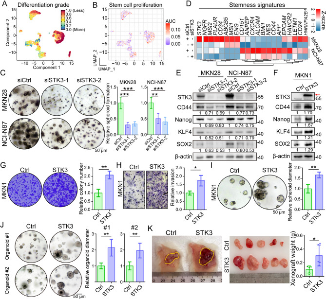
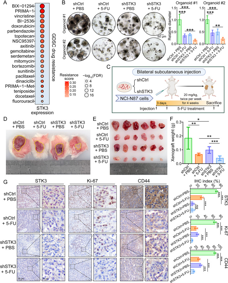
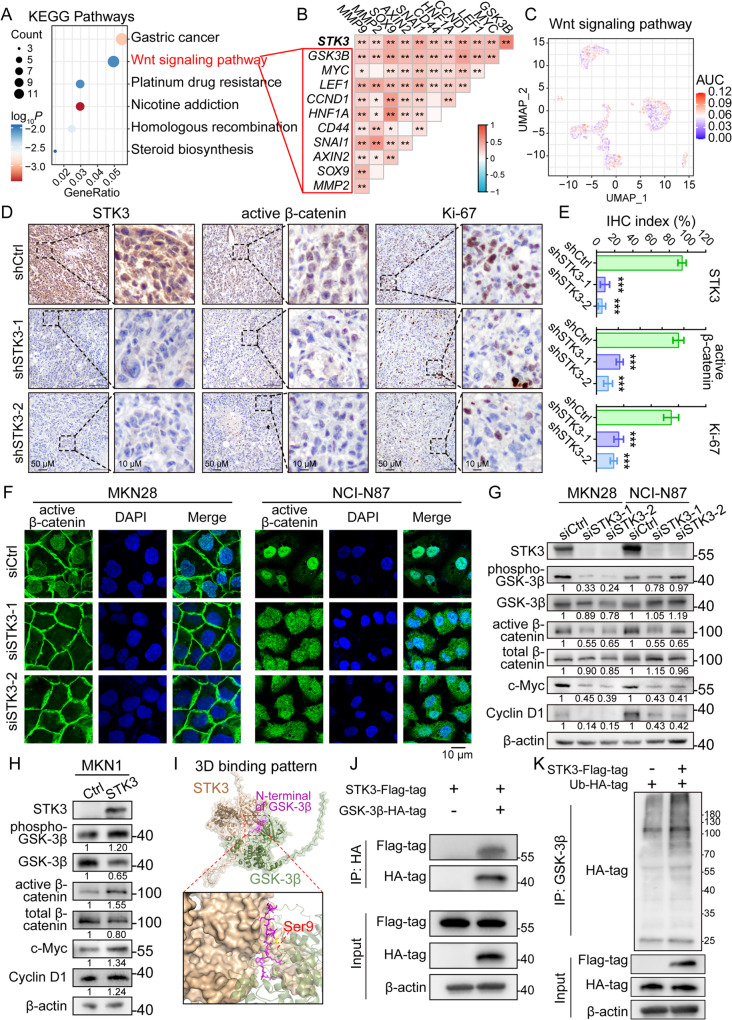
Results: Reduction of YAP1 significantly impaired STK3 expression at both mRNA and protein levels, and deletion of STK3 partially attenuated the oncogenic activity of YAP1. Notably, MNNG-induced tumors in Yap1-/-Taz-/- mice exhibited decreased STK3 expression. Knockdown of STK3 led to reduced expression of stemness markers and xenograft growth, while sensitizing GC organoids and xenografts to 5-fluorouracil treatment. Mechanistically, the direct interaction between STK3 and GSK-3β promoted GSK-3β phosphorylation and β-catenin nuclear accumulation, and thus the activation of Wnt signaling. Furthermore, aminopterin demonstrates as a promising STK3-targeted small molecule with remarkable effectiveness in inhibiting GC cell malignance and xenograft growth.

Conclusions: STK3 was identified as a transcriptional target of YAP1, leading to enhanced DNA repair ability and stemness acquisition during GC progression by activating Wnt/β-catenin activity through GSK-3β degradation. Moreover, STK3-targeted therapy offered a novel approach to concur acquired chemo-resistance in GC patients.

Keywords: Gastric cancer; STK3; Wnt signaling; YAP1.


Conflict of interest statement

Declarations. Ethical approval and consent to participate: The use of human samples was approved by the Joint Chinese University of Hong Kong-New Territories East Cluster Clinical Research Ethics Committee, Hong Kong (CREC Ref. No.: 2022-060). A waiver of consent form was granted by the Ethics Committee. Competing interests: The authors declare no competing interests.

Figures

Fig. 1
Fig. 1
STK3 is transcriptionally regulated by YAP1-TEAD4. (A) RNA-seq analysis based on YAP1-deleted cell lines. (B) Western bolt revealed the downregulation of STK3 protein expression level after YAP1 knockdown in MKN28 and NCI-N87. (C) STK3 level was upregulated after YAP1 overexpression in MKN1 and MKN7 cells. (D) A potential YAP1/TEAD4 binding motif was identified in STK3 promoter region. (E, F) YAP1-TEAD4 could directly bind to the promoter region of STK3 and exaggerate its expression, which is confirmed by public ChIP-seq dataset analysis and ChIP-qPCR assay. (G, H) Both VP and CA3 could downregulate the protein expression level of STK3. (I) Stk3 expression was significantly downregulated in Yap1−/−Taz−/− transgenic mice. (J, K) STK3 knockdown impaired the colony formation and invasion capacity of MKN28 and NCI-N87, while overexpressing YAP1 failed to rescue the detriment. (*, P < 0.05; **, P < 0.01; ***, P < 0.001)
Fig. 2
Fig. 2
STK3 shows co-upregulation with YAP1 in tumor tissues and cell lines across various GC cohorts. (A, B) Representative IHC images of YAP1-STK3 dual-negative and dual-positive GC primary samples. (C, D) Co-upregulation of YAP1 and STK3 was observed in Hong Kong cohort and proteome dataset STAD-CPYAC with a significant correlation. (E, F) The mRNA level of STK3 was significantly correlated with both YAP1 and TEAD4 expression in TCGA and ACRG cohort. (G) The mRNA expression of YAP1 and STK3 was remarkably associated in both all cancer cell lines and STAD cell lines. (H) UMAP reduction and cell type classification of a public GC scRNA-seq atlas. (I) The scRNA-seq data revealed that YAP1 and STK3 were largely overexpressed in cancer cells. (J-K) STK3+ and YAP1+ cancer cells were clustered in the same sub-clusters
Fig. 3
Fig. 3
STK3 gene is amplified in GC tissues, playing a crucial role in the regulation of cell cycle and DNA damage repair progression. (A) Copy number gain of STK3 gene was widely observed crossing all molecular subtypes of TCGA-STAD cohort (POLE, polymerase epsilon-mutated and ultramutated; MSI, microsatellite instability-high; GS, genomically stable; EBV Epstein-Barr virus-positive; CIN, chromosomal instability). (B) FISH assay demonstrated the frequent occurrence of STK3 gene copy number gain and amplification in primary GC samples. (C) The mRNA level of STK3 and YAP1 were remarkably correlated with the expression of cell cycle markers and DNA repair regulators in TCGA-STAD cohort. (D) The expression of cell cycling and DNA repair signatures are significantly upregulated in STK3+ samples when comparing with STK3 samples. (E) A volcano plot illustrating different expressed genes between STK3+ and STK3 samples. (F, G) Enrichment analysis and GSEA highlighted the hyperactivation of cell cycling- and DNA damage repair-related biological processes in STK3+ samples. (H, I) Pseudotime analysis on the scRNA-seq data indicated that STK3 was upregulated in the early stage of cancer cell evaluation, along with cell proliferation gene markers. (J, K) GSVA indicated a higher activity of DNA replication and DNA repair pathways in STK3+ cells. (L, M) RNA-seq analysis on STK3-deleted GC cell lines demonstrated declined expression of cell cycling/DNA repair-related gene expression and impaired pathway activities. (N) Phosphorylation of ATM, ATR, CHK1 and the expression of γ-H2AX were downregulated by STK3 depletion. (O) STK3 depletion impaired the xenograft formation capacity of NCI-N87 cells (n = 10 mice/group; ***, P < 0.001)
Fig. 4
Fig. 4
STK3 facilitates the acquisition of stemness features in GC cells, consequently accelerating tumor formation. (A, B) STK3+ cancer cells demonstrated less differentiation grade and higher activity in stem cell proliferation. (C) Knockdown of STK3 impaired the spheroid formation ability of MKN28 and NCI-N87. (D) STK3-depletion downregulated the mRNA levels of multiple stemness signatures. (E) Western blot assay indicated downregulated protein expressions of stemness markers in STK3-deleted cells, such as CD44, Nanog, KLF4, and SOX2. (F) Overexpression of STK3 increased the expression of CD44, Nanog, KLF4, and SOX2. (G-I) STK3 overexpression significantly enhanced the colony formation, invasion, and spheroid formation ability of MKN1 cells. (J, K) STK3 overexpression accelerated the growth of patient-derived organoids and cell line-derived xenografts. (n = 5 mice/group; *, P < 0.05; **, P < 0.01; ***, P < 0.001)
Fig. 5
Fig. 5
Knockdown of STK3 remarkably improves the GC chemotherapy sensitivity. (A) High STK3 expression level is correlated to the resistance of various chemotherapy drugs in GDSC dataset. (B) STK3 knockdown enhanced the sensitivity of GC organoids to 5-FU treatment, as indicated by the smaller organoid sizes in shSTK3 + 5-FU group. (C) Schematics of the in vivo drug sensitivity assay. (D) Representative photos of subcutaneous xenografts. (E, F) Xenograft formation was significantly restrained in STK3-deletion group and demonstrated heightened sensitivity to 5-FU treatment (n = 6 mice/group). (G) Representative IHC images of xenograft sections stained by STK3, cancer cell proliferation marker Ki-67, and stemness marker CD44 (ns, not significant; *, P < 0.05; **, P < 0.01; ***, P < 0.001)
Fig. 6
Fig. 6
STK3 mediates Wnt signaling transduction by directly interacting with GSK-3β. (A) Wnt signaling pathway was highlighted by enrichment analysis. (B) STK3 mRNA expression was positively correlated with multiple Wnt signaling signatures. (C) STK3 positive cells presented higher Wnt signaling activity. (D, E) Representative IHC images of xenograft sections stained by STK3, active β-catenin, and Ki-67. (F) Knockdown of STK3 inhibited the nuclear accumulation of active β-catenin. (G) STK3 depletion inhibited the phosphorylation of GSK-3β, leading to the upregulation of β-catenin degradation and downregulation of Wnt signaling signatures. (H) Overexpression of STK3 upregulated the phosphorylation of GSK-3β, and the expression of Wnt signaling markers. (I) The predicted binding pattern of STK3 and GSK-3β. (J) Co-IP assay proving the direct interaction between STK3 and GSK-3β. (K) introduction of STK3 accelerated the ubiquitination process of GSK-3β (***, P < 0.001)
Fig. 7
Fig. 7
Aminopterin is screened out by in silico modeling as a highly promising inhibitor for STK3. (A-B) Amin was identified as the most effective STK3 inhibitor among 4511 candidates with anti-tumor properties. (C-D) Predicted binding pattern and intermolecular interactions of Amin-STK3 complex (E) CETSA was conducted to further prove the binding between Amin and STK3. (F, G) Colony and spheroid formation ability of GC cell lines were restrained by Amin treatment in a dose-dependent manner. (H) Amin treatment downregulated the expression level of Wnt signaling signatures, cell cycling regulators, and stemness markers. (I) Amin administration impaired the growth of GC organoids and enhanced their sensitivity to 5-FU treatment (*, P < 0.05)

Similar articles

References

    1. Bray F, Laversanne M, Sung H, Ferlay J, Siegel RL, Soerjomataram I, Jemal A. Global cancer statistics 2022: GLOBOCAN estimates of incidence and mortality worldwide for 36 cancers in 185 countries. CA Cancer J Clin. 2024;74:229–63. - PubMed
    1. Smyth EC, Nilsson M, Grabsch HI, van Grieken NC, Lordick F. Gastric cancer. Lancet. 2020;396:635–48. - PubMed
    1. Li K, Zhang A, Li X, Zhang H, Zhao L. Advances in clinical immunotherapy for gastric cancer. Biochim Biophys Acta Rev Cancer. 2021;1876:188615. - PubMed
    1. Liu J, Yuan Q, Guo H, Guan H, Hong Z, Shang D. Deciphering drug resistance in gastric cancer: potential mechanisms and future perspectives. Biomed Pharmacother. 2024;173:116310. - PubMed
    1. Zheng Y, Pan D. The Hippo signaling pathway in development and disease. Dev Cell. 2019;50:264–82. - PMC - PubMed

MeSH terms

LinkOut - more resources