Artificial intelligence-assisted endobronchial ultrasound for differentiating between benign and malignant thoracic lymph nodes: a meta-analysis
- PMID: 40604844
- PMCID: PMC12224789
- DOI: 10.1186/s12890-025-03760-4
Artificial intelligence-assisted endobronchial ultrasound for differentiating between benign and malignant thoracic lymph nodes: a meta-analysis
Abstract
Background: Endobronchial ultrasound (EBUS) is a widely used imaging modality for evaluating thoracic lymph nodes (LNs), particularly in the staging of lung cancer. Artificial intelligence (AI)-assisted EBUS has emerged as a promising tool to enhance diagnostic accuracy. However, its effectiveness in differentiating benign from malignant thoracic LNs remains uncertain. This meta-analysis aimed to evaluate the diagnostic performance of AI-assisted EBUS compared to the pathological reference standards.
Methods: A systematic search was conducted across PubMed, Embase, and Web of Science for studies assessing AI-assisted EBUS in differentiating benign and malignant thoracic LNs. The reference standard included pathological confirmation via EBUS-guided transbronchial needle aspiration, surgical resection, or other histological/cytological validation methods. Sensitivity, specificity, diagnostic likelihood ratios, and diagnostic odds ratio (OR) were pooled using a random-effects model. The area under the receiver operating characteristic curve (AUROC) was summarized to evaluate diagnostic accuracy. Subgroup analyses were conducted by study design, lymph node location, and AI model type.
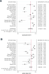
Results: Twelve studies with a total of 6,090 thoracic LNs were included. AI-assisted EBUS showed a pooled sensitivity of 0.75 (95% confidence interval [CI]: 0.60-0.86, I² = 97%) and specificity of 0.88 (95% CI: 0.83-0.92, I² = 96%). The positive and negative likelihood ratios were 6.34 (95% CI: 4.41-9.08) and 0.28 (95% CI: 0.17-0.47), respectively. The pooled diagnostic OR was 22.38 (95% CI: 11.03-45.38), and the AUROC was 0.90 (95% CI: 0.88-0.93). The subgroup analysis showed higher sensitivity but lower specificity in retrospective studies compared to prospective ones (sensitivity: 0.87 vs. 0.42; specificity: 0.80 vs. 0.93; both p < 0.001). No significant differences were found by lymph node location or AI model type.
Conclusion: AI-assisted EBUS shows promise in differentiating benign from malignant thoracic LNs, particularly those with high specificity. However, substantial heterogeneity and moderate sensitivity highlight the need for cautious interpretation and further validation.
Systematic review registration: PROSPERO CRD42025637964.
Keywords: Artificial intelligence; Endobronchial ultrasound; Malignancy; Meta-analysis; Thoracic lymph node.
© 2025. The Author(s).
Conflict of interest statement
Declarations. Ethics approval and consent to participate: Institutional Review Board approval was not required because this is a meta-analysis. Consent for publication: Not applicable. Competing interests: The authors declare no competing interests.
Figures


References
-
- Rueschhoff AB, Moore AW, Jasahui MRP. Lung Cancer Staging—A clinical practice review. J Respiration. 2024;4:50–61. - DOI
Publication types
MeSH terms
LinkOut - more resources
Full Text Sources
Medical

