

Three-dimensional finite element analysis of teeth displacement patterns under four anchorage designs for maxillary molar distalization using clear aligners: a real-case based simulation study
- PMID: 40604865
- PMCID: PMC12224400
- DOI: 10.1186/s12903-025-06375-7
Three-dimensional finite element analysis of teeth displacement patterns under four anchorage designs for maxillary molar distalization using clear aligners: a real-case based simulation study
Abstract
Objective: To explore the three-dimensional (3D) displacement patterns of maxillary molar distalization using clear aligners (CA) under four anchorage designs and provide clinical guidelines for optimal traction methods.
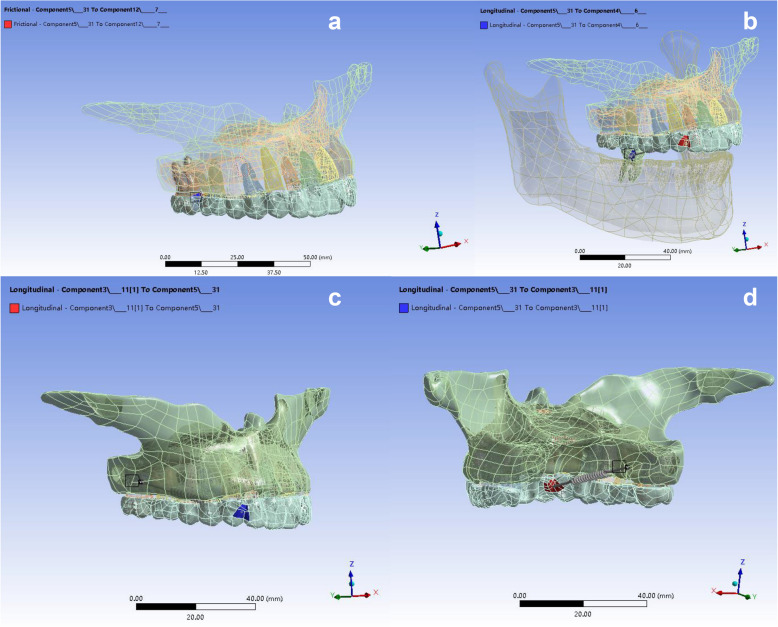
Materials and methods: A 3D finite element model was constructed based on CBCT and digital models from an adult patient requiring maxillary molar distalization. The model included cortical bone, cancellous bone, periodontal ligament, teeth, CA, and mini-screws. Four anchorage designs were simulated during sequential distal movement of bilateral maxillary second molars, first molars, and second premolars: (a) intramaxillary anchorage, (b) intermaxillary anchorage, (c) buccal mini-screw anchorage, and (d) palatal mini-screw anchorage. Displacement patterns of anterior and molar teeth were analyzed using ANSYS software and compared to the patient's actual maxillary dentition movement.
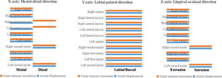
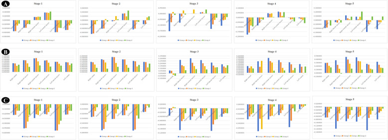
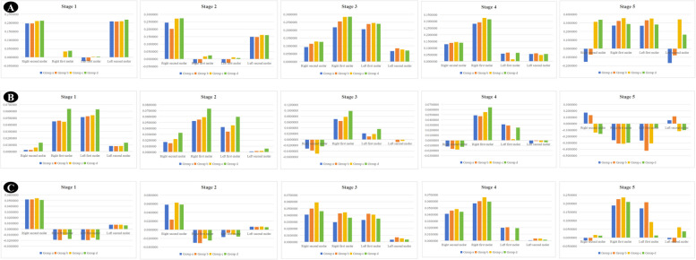
Results: Most teeth's actual displacement aligned with finite element predictions, except for central incisors, which showed mesial tipping instead of the simulated distal tipping. In the sequence-specific simulations: (1) Second molars only: All groups exhibited labial tipping and extrusion of anterior teeth (greater in Groups (a) and (b)), distal and buccal tipping with intrusion of second molars, and buccal tipping with extrusion of first molars (distal tipping in Groups (c) and (d), mesial in Groups (a) and (b)). (2) Second and first molars simultaneously: Anterior teeth showed reduced labial tipping, with palatal tipping and intrusion of right canines in Groups (c) and (d). All groups displayed distal tipping and intrusion of molars, with palatal tipping in second molars and buccal tipping in first molars. (3) Second premolars initiated: Anterior teeth in Groups (c) and (d) exhibited reduced labial tipping and extrusion, even with some palatal tipping and intrusion. Second molars in Groups (a) and (b) showed mesial, buccal tipping, and extrusion, while first molars in all groups and second molars in Groups (c) and (d) showed distal, palatal tipping, and intrusion. No significant differences in torque or vertical control were observed between buccal and palatal mini-screw anchorage.
Conclusions: Compared to intramaxillary or intermaxillary anchorage, the combination of mini-screws anchorage has a better effect on the torque control of anterior teeth, which also helps to promote the distal movement of molars. While no significant differences were observed in the torque and vertical control of anterior teeth or molars between buccal and palatal mini-screws, the findings underscore the versatility of mini-screw anchorage in accommodating individual patient anatomies. Notably, the displacement patterns of bilateral maxillary teeth were not always symmetrical, highlighting the need for personalized treatment planning and close monitoring during orthodontic therapy.
Keywords: Anchorage control; Clear aligners; Finite element analysis; Maxillary molar distalization.
© 2025. The Author(s).
Conflict of interest statement
Declarations. Ethics approval and consent to participate: Ethics approval for this study was obtained by the biomedical ethics committee of Peking University School and Hospital of Stomatology (PKUSSIRB No.202058139). The patient recruited in this study has signed informed consent forms before treatment. All the procedures were followed in accordance with the Declaration of Helsinki. Consent for publication: Not applicable. Competing interests: The authors declare no competing interests.
Figures
Similar articles
-
Biomechanical effects of clear aligners with different distal coverage designs combined with Class II elastic traction for maxillary first molar distalization: a finite element study.BMC Oral Health. 2025 Jul 2;25(1):1033. doi: 10.1186/s12903-025-06421-4. BMC Oral Health. 2025. PMID: 40604683 Free PMC article.
-
The effect of increasing the gaps between the front teeth on torque and intrusion control of the incisors for anterior retraction with clear aligners: a prospective study.BMC Oral Health. 2024 Jan 20;24(1):115. doi: 10.1186/s12903-024-03867-w. BMC Oral Health. 2024. PMID: 38243207 Free PMC article.
-
The effects of anterior spaces on the intrusion of mandibular incisors with clear aligners in extraction cases: a finite-element analysis.BMC Oral Health. 2025 Jul 4;25(1):1097. doi: 10.1186/s12903-025-06222-9. BMC Oral Health. 2025. PMID: 40615830 Free PMC article.
-
Maxillary molar distalization with noncompliance intramaxillary appliances in Class II malocclusion. A systematic review.Angle Orthod. 2008 Nov;78(6):1133-40. doi: 10.2319/101507-406.1. Angle Orthod. 2008. PMID: 18947282
-
Distalization of maxillary molars using temporary skeletal anchorage devices: A systematic review and meta-analysis.Orthod Craniofac Res. 2021 Mar;24 Suppl 1:103-112. doi: 10.1111/ocr.12470. Epub 2021 Feb 8. Orthod Craniofac Res. 2021. PMID: 33484608
References
-
- Liu X, Wang W, Gao J, Qin W, Wen Y, Luo H, Ma Y, Jin Z. Actual contribution ratio of maxillary and mandibular molars for total molar relationship correction during maxillary molar sequential distalization using clear aligners with Class II elastics: A finite element analysis. Am J Orthod Dentofacial Orthop. 2023;164(4):e106–20. - PubMed
MeSH terms
Grants and funding
LinkOut - more resources
Full Text Sources
