

The ultrasonographic evaluation of caudal vena cava diameter before and after fluid replacement in neonatal dehydrated calves with diarrhea
- PMID: 40604891
- PMCID: PMC12219989
- DOI: 10.1186/s12917-025-04759-z
The ultrasonographic evaluation of caudal vena cava diameter before and after fluid replacement in neonatal dehydrated calves with diarrhea
Abstract
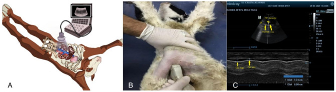
Background: In calves with diarrhea, it is critical to accurately determine the severity of dehydration and provide adequate fluid therapy. However, objective criteria are still limited. The aim of this study, a prospective cohort diagnostic study, is to compare caudal vena cava maximum diameter with expiration (CVCmax), caudal vena cava minimum diameter with inspiration (CVCmin), and caudal vena cava collapsibility index (CVC-CI) measurements before and after fluid therapy and to establish cut-off values for distinguishing between moderately and severely dehydrated calves. Twenty-four calves, with their degree of dehydration assessed based on enophthalmos and skin elasticity duration, were divided into two equal groups. Group I: consisted of 12 calves with an estimated degree of dehydration of 8-10% and were considered moderately dehydrated (degree of enophthalmos 4-6 mm, skin elasticity duration (s) 2-5). Group II: consisted of 12 calves with an estimated degree of dehydration 10-12% and were considered severely dehydrated (degree of enophthalmos 6-8 mm, skin elasticity duration (s) 5-10). Clinical examination, complete blood count and blood gas analysis, hemodynamic parameters (heart rate, respiratory rate, capillary refill time (CRT), L-lactate, systolic blood pressure (SBP), diastolic blood pressure (DBP), and mean arterial pressure (MAP)) and ultrasonographic examinations were performed for 48 h: before treatment (hour 0), immediately after the first fluid bolus, and at hours 8, 24, and 48 after the first fluid bolus. The Friedman test was used for within-group comparisons over time, and the Mann-Whitney U test was used for between-group comparisons at different time points. Categorical data were analysed using the chi-squared test, and Fisher's exact test was used when expected cell counts were less than 5. Receiver operating characteristic (ROC) analysis was performed to determine the sensitivity, specificity, and cut-off (lower limit) of CVC diameter and CVC-CI (%) compared with selected parameters (SBP, DBP, MAP, and L-lactate) to discriminate between moderate and severe dehydration. Statistical significance was set at P < 0.05.
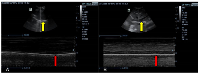
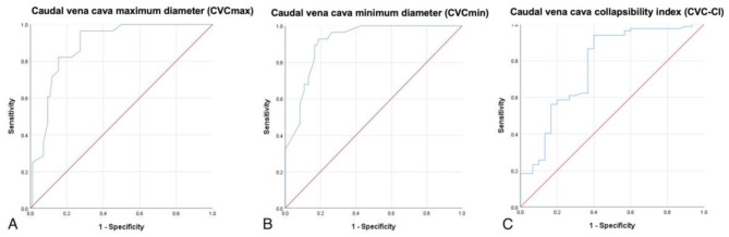
Results: CVCmax and CVCmin increased significantly after treatment in diarrheic calves (P < 0.05). Additionally, a significant decrease in CVC-CI (%) was observed in the treated diarrheic calves. Receiver operating characteristic (ROC) analysis showed that the area under the curve (AUC) of CVCmax was 0.885 (95% CI: 0.823-0.946; P < 0.001), with 82% sensitivity and 85% specificity at the intercept point of 1.05, the AUC of CVCmin was 0.913 (95% CI: 0.861-0.964; P < 0.001), with 89% sensitivity and 84% specificity at the intercept point of 0.66, and were the most reliable parameters in differentiating between moderate and severe dehydration.
Conclusion: A significant increase in CVCmax and CVCmin diameters, along with a significant decrease in CVC-CI, was observed with fluid therapy. The CVCmax and CVCmin diameters can provide valuable information for distinguishing between moderately and severely dehydrated calves.
Keywords: Calf; Caudal vena cava; Caudal vena cava collapsibility index; Dehydration; Neonatal diarrhea.
© 2025. The Author(s).
Conflict of interest statement
Declarations. Ethics approval and consent to participate: Approved by the Ethics Board of Selcuk University Veterinary Faculty Experimental Animals Production and Research Center (SUVDAMEK) Ethics Board (approval number: 2021/150). Every procedure was carried out in accordance with the relevant laws and standards. The study was conducted in compliance with the ARRIVE standards. The owner(s) of the animal gave their informed consent for us to use them in the study. Consent for publication: Not applicable. Competing interests: The authors declare no competing interests.
Figures
References
-
- Sen I, Constable PD. General overview to treatment of strong ion (metabolic) acidosis in neonatal calves with diarrhea. Eurasian J Vet Sci. 2013;29(3):114–20.
-
- Wilson M, Davis DP, Coimbra R, Via G, Tavazzi G, Price S, et al. Diagnosis and monitoring of hemorrhagic shock during the initial resuscitation of multiple trauma patients: a review. J Emerg Med. 2003;24(4):413–22. - PubMed
-
- Davis H, Jensen T, Johnson A. 2013 AAHA/AAFP fluid therapy guidelines for dogs and cats. J Am Anim Hosp Assoc. 2013;49(3):149–59. - PubMed
MeSH terms
Grants and funding
LinkOut - more resources
Full Text Sources
Medical
Research Materials
