. 2025 Jul 2;25(1):245.
doi: 10.1186/s12935-025-03853-4.

Combination of astragalus polysaccharide with Diosbulbin B exerts an enhanced antitumor effect in BRAFmut papillary thyroid cancer with decreased liver toxicity

Affiliations

Combination of astragalus polysaccharide with Diosbulbin B exerts an enhanced antitumor effect in BRAFmut papillary thyroid cancer with decreased liver toxicity

Shuai Xu et al. Cancer Cell Int. .

Abstract

Background: Diosbulbin B (DB) is a traditional Chinese medicine used for thyroid cancer treatment, but always brings severe liver injury. In the current study, we investigated the role of astragalus polysaccharide (APS) in DB-induced hepatotoxicity and their anti-tumor effect on BRAFmut papillary thyroid cancer (PTC), and disclosed the underlying mechanisms.

Methods: Two BRAFmut PTC IHH-4 and GLAG-66 cell lines were applied for the in vitro assays. CCK-8, flow cytometry, transwell chambers, enzyme-linked immunosorbent assay (ELISA) and transmission electron microscopy (TEM) were performed for cell growth, apoptosis, migration/invasion, malondialdehyde (MDA)/glutathione (GSH) content and mitochondria damage detection. Human normal liver epithelial cell line THLE-2 was used to assess the liver toxicity, together with the animal experiment.

Results: The IC50 of APS and DB in IHH-4 cells were 153.9 µg/mL and 41.2 µM, respectively, while they were 728.0 µg/mL and 22.74 µM in GLAG-66 cells. Combination of APS and DB enhanced the anti-cancer role of DB with increased cell apoptosis and LDH release, and weakened cell growth, migration and invasion capacities. Interestingly, the combination of these two drugs significantly alleviated the liver injury induced by DB. In mechanism, we found that APS combined with DB treatment triggered the increase of MDA level while decreased GSH level, and deteriorated mitochondria damage. Inhibition of ferroptosis impaired the anti-PTC role of APS combined with DB with no influencing on liver injury both in vivo and in vitro.

Conclusions: In conclusion, our study shows the combined therapy strategy of APS and DB regimen achieves better anti-cancer response through increasing MDA level while decreasing GSH level. Importantly, the combined therapy of APS and DB significantly decreased the liver toxicity induced by DB. These findings suggest that APS combined DB is a potential therapeutic strategy for BRAFmut PTC with high efficacy and low liver toxicity.

Keywords: BRAF mut papillary thyroid cancer; Astragalus polysaccharide; Diosbulbin B; Ferroptosis; Liver injury.


Conflict of interest statement

Declarations. Ethical approval: All animal experiments were performed in accordance with the institutional guidelines and approved by the Laboratory Animal Ethics Committee of Guangzhou University of Chinese Medicine (Approval no 20240221011). Consent for publication: Not applicable. Competing interests: The authors declare no competing interests.

Figures
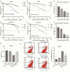
Fig. 1
Fig. 1
Combination of APS and DB induced PTC inhibition with decreased liver toxicity in vitro. The IC50 of APS (A) and DB (B) in IHH-4 cells was determined by CCK-8 test. (C) CCK-8 assay was used to evaluate the optimal concentration of DB in IHH-4 cells treated with 150 µg/mL APS. The IC50 of APS (D) and DB (E) in IHH-4 cells was determined by CCK-8 test. (F) CCK-8 assay was used to evaluate the optimal concentration of DB in IHH-4 cells treated with 700 µg/mL APS. (G) Cell viability was assessed using CCK-8 in the normal liver epithelial THLE-2 cells of the control, APS, DB and APS + DB groups. (H) Flow cytometry assay was performed to assess cell apoptosis in the normal liver epithelial THLE-2 cells of the control, APS, DB and APS + DB groups. (Three independent experiments were performed. *P<0.05 vs. control group; #P<0.05, vs. DB group, by the one-way analysis of variance with Tukey test)
Fig. 2
Fig. 2
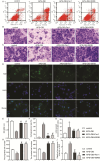
Combination of APS and DB significantly inhibited IHH-4 cell growth, migration and invasion. IHH-4 cells were submitted to different treatment, including APS (72 h), DB (72 h), APS + DB (72 h) and vemurafenib (24 h), and the following tests were carried out for different detections. (A, F) Cell apoptosis rate was determined using the flow cytometry assay. transwell chamber assay was used to evaluate cell migration (B, G) and invasion (C, H) abilities. (D) CCK-8 was used for cell growth assessment. (E) LDH release was detected to assess cytotoxicity. (Three independent experiments were performed, *P<0.05, vs. control group; #P<0.05, vs. DB group, by the one-way analysis of variance with Tukey test)
Fig. 3
Fig. 3
Combination of APS and DB significantly inhibited GLAG-66cell growth, migration and invasion. GLAG-66 cells were submitted to different treatment, including APS (72 h), DB (72 h), APS + DB (72 h) and vemurafenib (24 h), and the following tests were carried out for different detections. (A, F) Cell apoptosis rate was determined using the flow cytometry assay. transwell chamber assay was used to evaluate cell migration (B, G) and invasion (C, H) abilities. (D) CCK-8 was used for cell growth assessment. (E) LDH release was detected to assess cytotoxicity. (Three independent experiments were performed, *P<0.05, vs. control group; #P<0.05, vs. DB group, by the one-way analysis of variance with Tukey test)
Fig. 4
Fig. 4
Combination of APS and DB inhibited tumor growth with little liver toxicity in vitro. BALB/c nude mice bearing GLAG-66 cell-induced PTC tumors were treated with APS, DB, APS + DB and vemurafenib, following 28 days the tumor and liver tissues were collected for the subsequent assays, as follows. (A) Tumor images of the above group were made. (B, C) Tumor weight and volume were recorded. (D) HE staining was used to assess the live injury of mice with different treatment. (3 mice were included in each group, *P<0.05, vs. control group; #P<0.05, vs. DB group, by the one-way analysis of variance with Tukey test)
Fig. 5
Fig. 5
Combination of APS and DB activated the PERK/Nrf2/HO-1 signaling pathway in PTC cells. IHH-4 and GLAG-66 cells were submitted to different treatment, including APS (72 h), DB (72 h), APS + DB (72 h) and vemurafenib (24 h), and the following tests were carried out for different detections. The protein levels of COX2, eIF2a, GPX4, HO-1, PERK, Nrf2 and NCOA4 were detected using western blotting assay in (A) IHH-4 and (B) GLAG-66 cells. (C, D) GSH and (E, F) MDA levels were detected using ELISA. (G, H) TEM was used to assess mitochondria injury, red arrows indicate the mitochondria and green arrows indicate the mitochondrial cristae disruption (magnification: 10x). (Three independent experiments were performed, *P<0.05, vs. control group; #P<0.05, vs. DB group, by the one-way analysis of variance with Tukey test)
Fig. 6
Fig. 6
Inhibition of PERK/Nrf2/HO-1 or ferroptosis impaired the anti-tumor role of APS combined DB. GLAG-66 cells were treated with APS + DB, APS + DB + Fer-1 (an inhibitor of ferroptosis), or APS + DB + ISRIB (an inhibitor of eIF2α phosphorylation), and the following assays were carried out after treatment with the indicated time. (A and F) Flow cytometry assay was used to test cell apoptosis. Transwell chamber assay was used to evaluate (B and H) cell migration and (C and I) invasion abilities. (D and J) Immunofluorescence staining was used to assess the expression and location of Nrf2. (E) CCK-8 was used for cell viability detection. (G) LDH release was detected to assess cytotoxicity. (Three independent experiments were performed, *P<0.05, vs. control group; #P<0.05, vs. APS + DB group, by the one-way analysis of variance with Tukey test)
Fig. 7
Fig. 7
Inhibition of ferroptosis abandoned the anti-tumor role of combination of APS and DB in vitro. BALB/c nude mice bearing GLAG-66 cell-induced PTC tumors were treated with APS + DB, APS + DB + Fer-1, following 28 days the tumor and liver tissues were collected for the subsequent assays, as follows. (A) Tumor images of the above group were made. (B, C) Tumor weight and volume were recorded. (D, E) IHC staining was used to assess the expression of proliferative indexes (Ki-67) and PCNA in the tumor tissues of different groups (scale bar = 20 μm; magnification: 20x). (F) Western blotting was carried out to assess the protein expression levels of COX2, GPX4 and NCOA4 in the tumor tissues collected from mice with different treatments. (Three mice were included in each group, *P<0.05, vs. control group; #P<0.05, vs. APS + DB group, by the one-way analysis of variance with Tukey test)
Fig. 8
Fig. 8
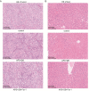
Morphology assessment of liver and tumor tissues. (A) HE staining was used to assess the tumor morphology. (B) HE staining was performed to assess live injury of mice with different treatment. (Three mice were included in each group)

Similar articles

References

    1. Zhang P, Guan H, Yuan S, Cheng H, Zheng J, Zhang Z, et al. Targeting myeloid derived suppressor cells reverts immune suppression and sensitizes BRAF-mutant papillary thyroid cancer to MAPK inhibitors. Nat Commun. 2022;13(1):1588. - PMC - PubMed
    1. Hong S, Xie Y, Cheng Z, Li J, He W, Guo Z, et al. Distinct molecular subtypes of papillary thyroid carcinoma and gene signature with diagnostic capability. Oncogene. 2022;41(47):5121–32. - PMC - PubMed
    1. Huang J, Wang J, Xv J, Wang J, Wang G, Zhao Y. Genetic alterations and allele frequency of BRAF V600E and TERT mutation in papillary thyroid carcinoma with intermediate-to-high recurrence risk: a retrospective study. Clin Exp Med. 2024;24(1):76. - PMC - PubMed
    1. Dadafarin S, Rodriguez TC, Carnazza MA, Tiwari RK, Moscatello A, Geliebter J. MEG3 expression indicates lymph node metastasis and presence of Cancer-Associated fibroblasts in papillary thyroid Cancer. Cells. 2022;11:19. - PMC - PubMed
    1. Li J, Zhang S, Zheng S, Zhang D, Qiu X. The BRAF V600E mutation predicts poor survival outcome in patients with papillary thyroid carcinoma: a meta analysis. Int J Clin Exp Med. 2015;8(12):22246–53. - PMC - PubMed

LinkOut - more resources