. 2025 Jul 2;16(1):347.
doi: 10.1186/s13287-025-04462-8.

LPS pretreated dental follicle stem cell derived exosomes promote periodontal tissue regeneration via miR-184 and PPARα-Akt-JNK signaling pathway

Affiliations

LPS pretreated dental follicle stem cell derived exosomes promote periodontal tissue regeneration via miR-184 and PPARα-Akt-JNK signaling pathway

Liangrui Chen et al. Stem Cell Res Ther. .

Abstract

Purpose: Lipopolysaccharide (LPS) pretreated dental follicle stem cells (DFSCs)-derived exosomes (L-D-Exo) exhibit enhanced therapeutic effects in periodontitis treatment, but the effective components responsible for these effects remain unidentified. The aim of this study is to investigate the differences in expression profile and regulatory effect of the exosomal microRNAs (miRNAs) from DFSCs and PDLSCs on periodontal tissue regeneration.

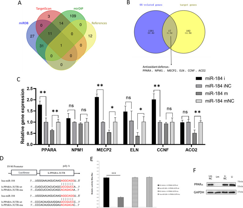
Methods: High-throughput miRNA sequencing was performed on DFSCs and PDLSCs derived exosomes under both Porphyromonas gingivalis (P.g) LPS pretreatment and normal conditions. Through bioinformatic analysis, miR-184 was selected as the key miRNA due to its specific down-regulation in L-D-Exo, which linked to oxidative stress regulation. After changing the expression of miR-184 in PDLSCs, the fluorescence intensity of reactive oxygen species (ROS), malondialdehyde (MDA) content and antioxidant related enzyme activities, and the expression levels of inflammatory cytokines and osteogenesis-related genes in PDLSCs were detected. In addition, dual-luciferase reporter assay and Western blot were used to explore the target gene and downstream signaling pathways. In vivo, miR-184 Antagomir was injected into mice with experimental periodontitis to evaluate the role and mechanism of miR-184 in periodontal tissue regeneration.

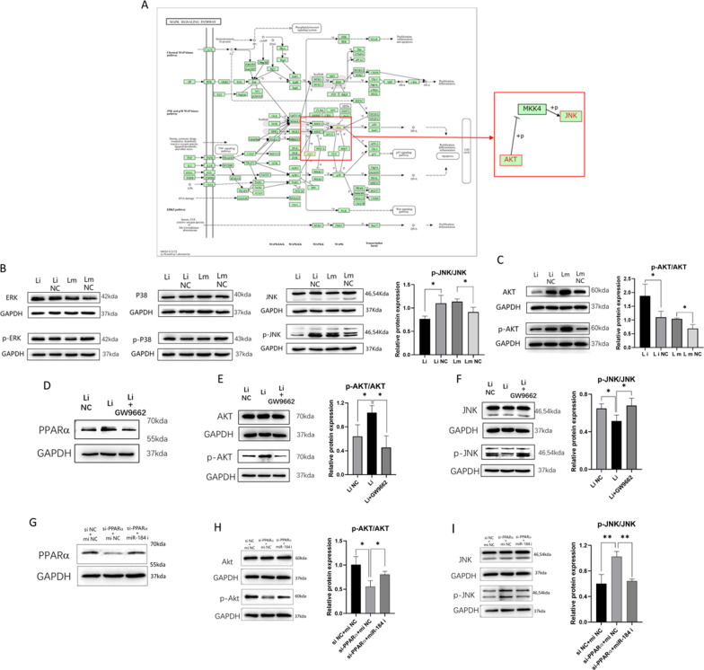
Results: Inhibition of miR-184 in PDLSCs significantly impaired oxidative stress, as evidenced by decreased ROS fluorescence intensity and MDA content, alongside increased activities of antioxidant enzymes. This reduction in oxidative stress subsequently decreased the expression of intracellular inflammatory cytokines, while promoting the expression of osteogenic genes. The dual-luciferase reporter assay confirmed the direct binding of miR-184 with Peroxisome proliferator-activated receptor α (PPARα). MiR-184 inhibition activated the downstream protein kinase B (Akt) pathway and inhibited the c-Jun N-terminal kinase (JNK) pathway under inflammatory conditions. Furthermore, miR-184 Antagomir application also enhanced the therapeutic efficacy of periodontitis mice by reducing inflammation and promoting periodontal osteogenesis.

Conclusion: Inhibition of miR-184 facilitates periodontal regeneration, which targets the PPARα-Akt-JNK signaling pathway to suppress oxidative stress in periodontal tissues.

Keywords: Dental follicle stem cells; Exosomes; MicroRNA; Oxidative stress; Periodontal tissue regeneration.


Conflict of interest statement

Declarations. Ethics approval and consent to participate: Informed consent was obtained from all patients under ethical approval (WCHSIRB-D-2023-165, titled “Dental follicle stem cell-derived exosomes promote periodontal tissue regeneration via miR-184 and its mechanisms”) by the Ethics committee of West China Hospital of Stomatology (date of approval: November 16, 2023) before utilizing donated dental specimens for further study. The animal study was performed according to the Institutional Animal Care and Use Committee (IACUC) at Sichuan University, and approved (WCHSIRB-D-2023-037, titled “Dental follicle stem cell-derived exosomes promote periodontal tissue regeneration via miR-184 and its mechanisms”) by the Ethics Committee of West China Hospital of Stomatology (date of approval: February 16, 2023). Consent for publication: Not applicable. Competing interests: The authors declare that they have no competing interests.

Figures

Fig. 1
Fig. 1
Characteristics of exosomes. A Flowchart of stem cells and exosomes isolation. B Transmission electron micrograph of exosomes. C Exosome particle size distribution analyzed by nanoparticle tracking. D The detection of exosomal surface markers through western blot, full-length blots are presented in Supplementary Fig. 2A. E Photographs of colocalization of PDLSCs and exosomes under confocal microscopy
Fig. 2
Fig. 2
Differential expression profiling of exosomal miRNAs in DFSCs and PDLSCs under normal and microinflammatory conditions. A Heat maps depicted differential miRNAs of L-D-Exo compared to D-Exo and L-P-Exo compared to P-Exo, with red-white-blue color indicating the miRNA expression from high to low in turn. B Histograms demonstrated the top ten bars of GO enrichment analysis. C Bubble plots showed the top ten pathways that the predicted target genes were enriched to in each group. D Heat maps depicted differential miRNAs of D-Exo compared to P-Exo and L-D-Exo compared to L-P-Exo, with red–black–green color indicating the miRNA expression from high to low in turn. E Histograms showed GO enrichment analysis in D-Exo compared to P-Exo and L-D-Exo compared to L-P-Exo. F Bubble plots showed KEGG enrichment analysis in D-Exo compared to P-Exo and L-D-Exo compared to L-P-Exo, in which MAPK signaling pathway and PI3K-Akt signaling pathway were repeatedly enriched in
Fig. 3
Fig. 3
Sequencing results validation and key miRNA screening. A qRT-PCR verification of differential miRNAs in exosomes of DFSCs after LPS pretreatment. B QRT-PCR verification of differential miRNAs in exosomes of PDLSCs after LPS pretreatment. C Four sets of miRNA sequencing data and analysis methods, including vertical (shown by blue arrows) and horizontal analysis (shown by red arrows). D Venn diagram showed the intersection of differential miRNAs in vertical and horizontal analysis
Fig. 4
Fig. 4
Oxidative stress, inflammatory response and osteogenesis of PDLSCs after miR-184 transfection. A, B ROS fluorescence microscope photograph of PDLSCs with nuclei in blue and ROS fluorescence in green. Semi-quantitative results were presented. C Concentration of MDA. D Antioxidant SOD enzyme activity. E Antioxidant CAT enzyme activity. F Antioxidant GSR enzyme activity. G Gene expression levels of inflammatory cytokines IL-6, IL-1β and TNF-α. H The expression levels of osteogenesis-related genes OCN, ALP, POSTN and RUNX2
Fig. 5
Fig. 5
MiR-184 target gene screening and validation. A Venn diagram showed all miR-184 target genes predicted in MiRDB, TargetScan and mirDIP database and reported in the literature searched on PubMed. B The intersection of miR-184 target genes and 1398 oxidative stress-related genes was analyzed, among which PPARA, NPM1, MECP2, ELN, CCNF and ACO2 were involved in oxidative stress defense. C The gene expression of PPARA, NPM1, MECP2, ELN, CCNF and ACO2 after miR-184 transfection verified by qRT-PCR. D Target site of hsa-miR-184 binding to h-PPARA-3UTR-WT and the mutation sites of h-PPARA-3UTR-MUT. E Hsa-miR-184 significantly down-regulated h-PPARA-3UTR-WT luciferase expression and failed to down-regulate luciferase expression of h-PPARA-3UTR-MUT. F MiR-184 downregulated PPARα protein expression, while inhibition of miR-184 promoted PPARα protein expression, full-length blots are presented in Supplementary Fig. 2B
Fig. 6
Fig. 6
Exploration of miR-184 downstream signaling pathway. A The position of Akt and JNK (marked in red) in the MAPK signaling pathway network demonstrated by KEGG. B Changes in ERK, JNK, and P38 signaling pathways after miR-184 transfection, in which the JNK signaling pathway was inhibited after transfection with miR-184 inhibitor. C Changes in AKT signaling pathway after miR-184 transfection and efficiency of AKT activation was increased by transfection with miR-184 inhibitor. D PPARα protein expression was down-regulated after transfection with miR-184 inhibitor and addition of GW9662. E The Akt signaling pathway was inhibited after transfection with miR-184 inhibitor and suppression of PPARα expression. F The JNK signaling pathway was activated after transfection with miR-184 inhibitor and suppression of PPARα expression. G-I PPARα, Akt and JNK pathway protein expression changes after co-transfection with si-PPARα and miR-184 inhibitor. The above full-length blots are presented in Supplementary Fig. 2C-J
Fig. 7
Fig. 7
Application of miR-184 Antagomir in periodontitis mice. A Micro-CT images of mice maxilla at 2 weeks post-treated. Scale bar: 1 mm. The red box represents the area of periodontal defect. B Statistical analysis of CEJ-ABC distance in mesial buccal root of maxillary second molar. C The representative H&E and Masson stained sections. Scale bar: 100 μm. D The representative TRAP stained sections. Scale bar: 100 μm. E Quantitative analysis of TRAP staining. F–G The IHC staining images of RANKL, OCN, POSTN, OPG, TNF-α, IL-1β and their expression quantitative analysis. Scale bar: 200 μm. D, cementum; PL, periodontal ligament; AB, alveolar bone. H–I The IF staining images of PPARα and DAPI and their Fluorescence intensity analysis. Scale bar: 50 μm

Similar articles

References

    1. Frencken JE, Sharma P, Stenhouse L, Green D, Laverty D, Dietrich T. Global epidemiology of dental caries and severe periodontitis—a comprehensive review. J Clin Periodontol. 2017;44(Suppl 18):S94-s105. - PubMed
    1. Slots J. Periodontitis: facts, fallacies and the future. Periodontology 2000. 2017;75(1):7–23. - PubMed
    1. Hu L, Liu Y, Wang S. Stem cell-based tooth and periodontal regeneration. Oral Dis. 2018;24(5):696–705. - PubMed
    1. Xu XY, Li X, Wang J, He XT, Sun HH, Chen FM. Concise review: periodontal tissue regeneration using stem cells: strategies and translational considerations. Stem Cells Transl Med. 2019;8(4):392–403. - PMC - PubMed
    1. Guo S, Kang J, Ji B, Guo W, Ding Y, Wu Y, et al. Periodontal-derived mesenchymal cell sheets promote periodontal regeneration in inflammatory microenvironment. Tissue Eng Part A. 2017;23(13–14):585–96. - PubMed

MeSH terms