

Mature retroperitoneal cystic teratoma in an adult alpaca: a case report
- PMID: 40605025
- PMCID: PMC12219530
- DOI: 10.1186/s12917-025-04875-w
Mature retroperitoneal cystic teratoma in an adult alpaca: a case report
Abstract
Background: Retroperitoneal mature cystic teratomas are rare in both human and veterinary medicine. These tumors arise from pluripotent germ cells and can contain tissues from all three germ layers. While the majority of teratomas are benign, their location in the retroperitoneum can lead to significant mass effects and clinical complications.
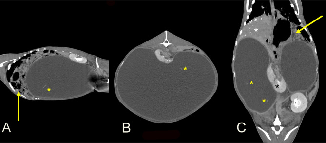
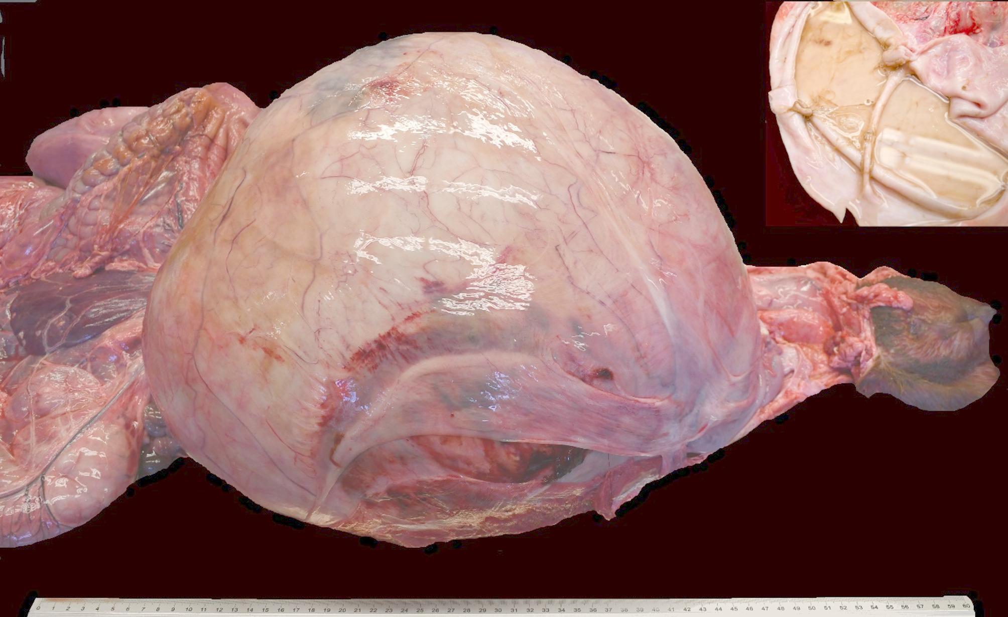
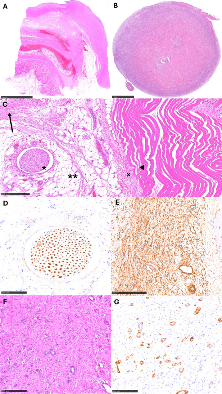
Case presentation: A four-year-old female alpaca was presented with a history of abdominal distension. Clinical signs included tenesmus with lifted tail, reduced fecal output and hypothermia. Initial ultrasound revealed severe accumulation of anechoic fluid in the abdomen and complete homogeneous soft tissue opacification of the abdomen was visible in radiography, with mass effect on all other abdominal organs. A CT scan confirmed a considerable cystic lesion in the caudal abdomen. Histopathological analysis of the cystic structure revealed mature connective tissue, both striated and smooth muscle, as well as gland-like formations. Immunohistochemical staining identified markers from multiple tissue types. Based on these findings, a diagnosis of mature retroperitoneal cystic teratoma was established.
Conclusions: To our knowledge, this is the first reported case of a mature retroperitoneal cystic teratoma in an adult alpaca. The rare nature of this condition in both humans and animals makes it a noteworthy addition to veterinary literature. This case emphasizes the importance of considering such conditions in the differential diagnosis of abdominal masses in livestock.
Keywords: Alpaca (vicugna pacos); Cystic teratoma; Histopathology; Radiology; Retroperitoneum.
Conflict of interest statement
Declarations. Ethics approval and consent to participate: Not applicable. Consent for publication: Informed consent was obtained from the animal’s owner for the publication of any data or images related to this case. Competing interests: The authors declare no competing interests.
Figures
References
-
- Meuten DJ, editor. Tumors in domestic animals. Wiley 2016.
-
- Hill FI, Mirams CH. Intracranial teratoma in an alpaca (Vicugna pacos) in new Zealand. Vet Rec. 2008;162:188–9. - PubMed
-
- Mutinelli F, Carminato A, Bozzato E, Marchioro W, Trevisan L, Vascellari M. Retroperitoneal teratoma in a domestic rabbit (Oryctolagus cuniculus). J Vet Med Sci. 2009;71:367–70. - PubMed
-
- Munday JS, Fairchild SE, Brown CA. Retroperitoneal teratoma in a skunk (Mephitis mephitis). J Zoo Wildl Med. 2004;35:406–8. - PubMed
LinkOut - more resources
Full Text Sources
