. 2025 Dec;30(1):2527427.
doi: 10.1080/13510002.2025.2527427. Epub 2025 Jul 2.

Ultraviolet B-induced oxidative damage in human skin keratinocytes is alleviated by Pinus morrisonicola leaf essential oil through activation of the Nrf2-dependent antioxidant defense system

Affiliations

Ultraviolet B-induced oxidative damage in human skin keratinocytes is alleviated by Pinus morrisonicola leaf essential oil through activation of the Nrf2-dependent antioxidant defense system

Wan-Teng Lin et al. Redox Rep. 2025 Dec.

Abstract

Background: Ultraviolet B (UVB) radiation contributes to skin disorders such as photodamage, photoaging, and cancer. Natural antioxidants can mitigate UVB-induced damage. Pinus morrisonicola (Taiwan white pine), known for its anti-cancer, anti-inflammatory, and antioxidant properties, is used in health-promoting beverages, but its skin-protective effects remain underexplored.

Purpose: This study investigates the protective effects of P. morrisonicola leaf essential oil (PMLEO) against UVB-induced damage in HaCaT keratinocytes.

Methods: HaCaT cells were exposed to UVB and treated with PMLEO. Cell viability, reactive oxygen species (ROS) levels, and antioxidant enzyme expression were assessed. The role of Nrf2, a key antioxidant regulator, was evaluated through knockdown experiments. The effects on UVB-induced melanogenesis were examined via α-MSH secretion followed by p53-mediated POMC expression.

Results: PMLEO and P. morrisonicola bark essential oil (PMBEO) were non-cytotoxic up to 200 µg/mL. UVB reduced cell viability to 43%, but PMLEO co-treatment significantly restored viability and reduced ROS levels via Nrf2 activation, increasing NQO-1 and HO-1. Nrf2 knockdown impaired PMLEO's protection. PMLEO also inhibited UVB-induced α-MSH secretion by downregulating p53-mediated POMC expression, suggesting an anti-melanogenic effect.

Conclusion: PMLEO protects dermal keratinocytes against UVB-induced oxidative stress, cell death, and melanogenesis via Nrf2 activation, highlighting its potential as a natural skin protectant.

Keywords: HaCaT; Nrf2 pathway; Pinus morrisonicola; antioxidant; essential oil; photoaging; photodamage; ultraviolet B.


Conflict of interest statement

No potential conflict of interest was reported by the author(s).

Figures

Figure 1.
Figure 1.
Cytotoxic effect of PMLEO, PMBEO, and UVB on HaCaT cells. (A) HaCaT cells were incubated with increasing concentrations of PMLEO (25–200 µg/mL) for 24 h. (B) HaCaT cells were incubated with increasing concentrations of PMBEO (25–200 µg/mL) for 24 h. (C) HaCaT cells were pre-treated with indicated concentrations of PMLEO and PMBEO for 2 h and then exposed to UVB (50 mJ/cm2) for 24 h. (D) HaCaT cells were pre-treated with various concentrations of PMLEO (25–100 µg/mL) for 2 h and then exposed to UVB (50 mJ/cm2) for 24 h. (E) HaCaT cells were pre-treated with various concentrations of PMBEO (25–100 µg/mL) for 2 h and then exposed to UVB (50 mJ/cm2) for 24 h. The cell viability was measured by MTT assay. The percentage of cell viability was compared with the control (0.01% DMSO) group. Values represent the mean ± SD of three independent experiments. Statistical significance was set at ΦP < 0.05 compared to control vs. UVB (50 mJ/cm2) and *P < 0.05, **P < 0.01, ***P < 0.001 compared with UVB alone treatment group vs. UVB + sample treatment groups.
Figure 2.
Figure 2.
PMLEO inhibits UVB-induced oxidative stress in HaCaT cells. (A) PMLEO inhibits UVB-induced intracellular ROS generation in HaCaT cells. (B) The free-radical scavenging activity of PMLEO was determined by DPPH assay. Ascorbic acid (AA) was used as positive control. Values represent the mean ± SD of three independent experiments. Statistical significance was set at ΦP < 0.05 compared to control vs. UVB (50 mJ/cm2) and **P < 0.01, ***P < 0.001 compared with UVB alone vs. UVB + sample treatment groups.
Figure 3.
Figure 3.
PMLEO prevents UVB-induced oxidative stress in HaCaT cells by employing endogenous anti-oxidants. (A,B) To quantify the mRNA expression level of HO-1 and NQO-1 HaCaT cells were pre-incubated with PMLEO (25–100 µM) or resveratrol (40 µM) and then exposed to UVB (50 mJ/cm2) for 6 h. Total RNA was extracted and subjected to Q-PCR analysis. Relative HO-1 and NQO-1 mRNA level were normalized with GAPDH mRNA. (C) To determine the protein expression levels of HO-1, NQO-1, and γ-GCLC, HaCaT cells were pre-incubated with PMLEO (25–100 µM) for 24 h. Total cell lysates were prepared and subjected to western blot analysis. Histogram shows the relative protein expression levels of HO-1, NQO-1, and γ-GCLC, which are normalized with an internal control GAPDH. Values represent the mean ± SD of three independent experiments. Statistical significance was set at ϕP < 0.05, ϕϕP < 0.01, ϕϕϕP < 0.001 compared with control a vs. sample treatment groups. *P < 0.05, **P < 0.01, ***P < 0.001 compared with UVB alone vs. UVB + sample treatment groups.
Figure 4.
Figure 4.
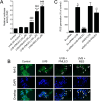
PMLEO up-regulates antioxidants via Nrf2 pathway. (A) To determine the Nrf2 transcriptional activity, HaCaT cells were transiently transfected with ARE promoter construct using lipofectamine and pre-incubated with PMLEO (25–100 µM) or resveratrol (40 µM) for 2 h and then exposed to UVB (50 mJ/cm2) for 6 h. Cell lysates were mixed with luciferase reagents and quantified using an illuminometer. Relative ARE promoter activity was calculated by dividing treated cells' relative luciferace unit (RLU) by RLU of untreated cells (control). (B) To determine the nuclear localization of Nrf2, HaCaT cells were pre-incubated with PMLEO (100 µg/mL) or resveratrol (40 µM) and then exposed to UVB (50 mJ/cm2) for 2 h. The protein expression and localization of Nrf2 was measured by immunofluorescence using Nrf2 specific primary antibody with FITC-conjugated secondary antibody (green). DAPI (1 µM) was used to stain the nucleus. (C) HaCaT cells were transfected with specific siRNA against Nrf2 or control siRNA. After transfection for 24 h, cells were pre-incubated with PMLEO (100 µM) for 2 h and then exposed to UVB (50 mJ/cm2) for 1 h. Intracellular ROS was measured by DCFH2-DA assay. Values represent the mean ± SD of three independent experiments. Statistical significance was set at ϕP < 0.05, ϕϕP < 0.01, ϕϕϕP < 0.001 compared with control a vs. sample treatment groups and **P < 0.01, ***P < 0.001 compared with UVB alone vs. UVB + sample treatment groups. ΔP < 0.05 compared to control siRNA + UVB vs. siNrf2 + UVB. ωP < 0.05 compared with siNrf2 + UVB vs. siNrf2 + UVB + PMLEO treatment group.
Figure 5.
Figure 5.
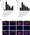
PMLEO suppresses UVB-induced α-MSH secretion by inhibiting p53 activation in HaCaT cells. (A) The intracellular levels of α-MSH were measured using a commercially available EIA kit. (B) To assess the mRNA expression of POMC, HaCaT cells were pre-treated with PMLEO (25–100 µM) or RES (40 µM), then exposed to UVB (50 mJ/cm2), and incubated for 6 h. Total RNA was extracted and analyzed via q-PCR, with POMC mRNA levels normalized to GAPDH mRNA. (C) The protein expression and localization of phosphorylated p53 (phos-p53) were evaluated by immunofluorescence using a phos-p53 specific primary antibody and a FITC-conjugated secondary antibody (purple). DAPI staining was used for nuclear visualization. Data are presented as mean ± SD from three independent experiments. Statistical significance was defined as ϕP < 0.05 and ϕϕϕP < .001for control versus UVB (50 mJ/cm2) and *P < 0.05, ***P < 0.01, ***P < 0.001 for UVB alone versus UVB with sample treatment. HaCaT refers to human skin keratinocytes, POMC to proopiomelanocortin, q-PCR to quantitative PCR, UVB to ultraviolet B, and α-MSH to α-melanocyte-stimulating hormone.

References

    1. Marrot L, Meunier JR.. Skin DNA photodamage and its biological consequences. J Am Acad Dermatol. 2008;58(5):S139–S148. doi: 10.1016/j.jaad.2007.12.007 - DOI - PubMed
    1. Gilchrest BA . Photoaging. J Invest Dermatol. 2013;133(E1):E2–E6. - PubMed
    1. Song X, Mosby N, Yang J, et al. alpha-MSH activates immediate defense responses to UV-induced oxidative stress in human melanocytes. Pigment Cell Melanoma Res. 2009;22(6):809–818. doi: 10.1111/j.1755-148X.2009.00615.x - DOI - PubMed
    1. Wagener FA, Carels CE, Lundvig DM.. Targeting the redox balance in inflammatory skin conditions. Int J Mol Sci. 2013;14(5):9126–9167. doi: 10.3390/ijms14059126 - DOI - PMC - PubMed
    1. Masaki H, Izutsu Y, Yahagi S, et al. Reactive oxygen species in HaCaT keratinocytes after UVB irradiation are triggered by intracellular Ca(2+) levels. J Investig Dermatol Symp Proc. 2009;14(1):50–52. doi: 10.1038/jidsymp.2009.12 - DOI - PubMed

MeSH terms