Using Dynamic Joint Space During Physiological Loading to Objectively Measure Hip Stability
- PMID: 40605408
- PMCID: PMC12329620
- DOI: 10.1002/jor.70011
Using Dynamic Joint Space During Physiological Loading to Objectively Measure Hip Stability
Abstract
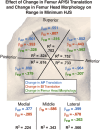
An objective measurement of hip stability during functional loading is needed to improve diagnosis, guide treatment decisions, and evaluate intervention success. This study aimed to develop a reference measurement for stable hips based upon dynamic minimum hip joint space (HJS) in healthy young adults. Synchronized biplane radiographs of the hips of 24 healthy young adults were collected (50 images/s) during treadmill walking and squatting. Subject-specific femur and pelvis models were created from CT scans, and bone motion was determined by a validated volumetric model-based tracking technique. The distance between femur and acetabulum subchondral bone surfaces was calculated during each movement. Range in minimum subchondral bone distance was measured in radial and sagittal regions of the acetabula. Regression analysis identified kinematics and morphologic predictors of range in minimum HJS. Range in minimum HJS during gait in the anterior-inferior (1.8 mm) and posterior-superior regions (1.7 mm) was 31%-38% larger than in the anterior-superior and superior regions (1.3 mm; p ≤ 0.001), and 13%-20% larger than in the posterior-inferior region (1.5 mm; p ≤ 0.001). No differences in minimum HJS were identified in radial regions during squat (range: 0.7-0.9 mm). No sex differences were identified. Femur head translation during gait was a stronger predictor of the range in minimum HJS than changes in femur head morphology. This suggests anterior-inferior to posterior-superior pistoning of the femur may be a mechanical mechanism for commonly observed pathology. These results suggest that gait is a better activity than squatting to assess dynamic hip stability when using this metric.
Keywords: dynamic biplane radiography; dynamic hip joint space; hip stability.
© 2025 The Author(s). Journal of Orthopaedic Research® published by Wiley Periodicals LLC on behalf of Orthopaedic Research Society.
Conflict of interest statement
Craig Mauro is a consultant for and receives royalties from Arthrex Inc. and Michael McClincy receives royalties from Elizur for an unrelated project. All other authors have no professional or financial affiliations that may be perceived to have biased the presentation.
Figures


References
-
- Martin H. D., Kelly B. T., Leunig M., et al., “The Pattern and Technique in the Clinical Evaluation of the Adult Hip: The Common Physical Examination Tests of Hip Specialists,” Arthroscopy 26 (2010): 161–172. - PubMed
-
- Reiman M. P., Goode A. P., Hegedus E. J., Cook C. E., and Wright A. A., “Diagnostic Accuracy of Clinical Tests of the Hip: A Systematic Review With Meta‐Analysis,” British Journal of Sports Medicine 47 (2013): 893–902. - PubMed
-
- Truntzer J. N., Hoppe D. J., Shapiro L. M., and Safran M. R., “Can the FEAR Index be Used to Predict Microinstability in Patients Undergoing Hip Arthroscopic Surgery?,” American Journal of Sports Medicine 47 (2019): 3158–3165. - PubMed
-
- Brusalis C. M., Peck J., Wilkin G. P., et al., “Periacetabular Osteotomy as a Salvage Procedure: Early Outcomes in Patients Treated for Iatrogenic Hip Instability,” Journal of Bone and Joint Surgery 102 (2020): 73–79. - PubMed
-
- Safran M. R., Murray I. R., Andrade A. J., et al., “Criteria for the Operating Room Confirmation of the Diagnosis of Hip Instability: The Results of an International Expert Consensus Conference,” Arthroscopy 38 (2022): 2837–2849.e2. - PubMed
MeSH terms
Grants and funding
LinkOut - more resources
Full Text Sources

