. 2025 Jul 8;166(9):bqaf115.
doi: 10.1210/endocr/bqaf115.

NEDD4 Promotes Sertoli Cell Proliferation and Adult Leydig Cell Differentiation in the Murine Testis

Affiliations

NEDD4 Promotes Sertoli Cell Proliferation and Adult Leydig Cell Differentiation in the Murine Testis

Simon Peter Windley et al. Endocrinology. .

Abstract

Successful testis development relies on the coordinated differentiation and assembly of various cell types to establish both endocrine and reproductive functions. The ubiquitin ligase NEDD4 has emerged as a key player in murine testis development, with this enzyme being implicated in gonadal sex determination and spermatogonial stem cell differentiation. Here, we report hitherto uncharacterized roles of NEDD4 in postnatal testis development. Utilizing Nr5a1- and Amh-Cre drivers to conditionally ablate Nedd4 in testicular somatic cells, we show that NEDD4 promotes Sertoli cell proliferation through the modulation of the PI3K-AKT signaling pathway. This ubiquitin ligase also ensures proper differentiation of adult Leydig cells and may contribute to murine steroidogenesis. Furthermore, NEDD4 is essential for adrenal gland differentiation, as its loss results in adrenal dysgenesis. These findings highlight NEDD4 as a crucial factor in testis development, emphasizing the importance of ubiquitination and post-translational modifications in reproductive biology.

Keywords: NEDD4; adrenal dysgenesis; steroidogenesis; testis development; testis function.


Figures

Figure 1.
Figure 1.
The testis-determining program is unaffected in XY Nr5a1-Cre;Nedd4Flox/Flox mice. Section immunofluorescence on 11.5 dpc (A) and 12.5 dpc (B) XY Nr5a1-Cre;Nedd4Flox/Flox gonads (bottom) alongside Cre-negative XY littermate controls (top) stained for Sertoli cell markers SRY (green, left panel), SOX9 (green, middle panel), and AMH (magenta right panel in B), granulosa cell marker FOXL2 (green, right panel) and germ cell marker DDX4 (magenta in A and left and middle panels of B). The anterior pole of each gonad is positioned at the top of each panel. Scale bars = 100 μm.
Figure 2.
Figure 2.
Unperturbed fetal testis differentiation in XY Nr5a1-Cre;Nedd4Flox/Flox mice. Section immunofluorescence on 14.5 dpc XY Nr5a1-Cre;Nedd4flox/flox testes alongside XY littermate controls stained for Sertoli cell markers SOX9 (green in A, E), GATA4 (green in B, G), and AMH (magenta in D, I), extracellular matrix protein laminin (magenta in B, G), Leydig cell marker CYP11A1 (green in C, H), germ cell marker DDX4 (magenta in A, E, F, J), pluripotency marker POU5F1 (magenta in C, H), meiosis marker SYCP3 (green in D, I), and granulosa cell marker FOXL2 (green in E, J). The anterior pole of each gonad is positioned at the top of each panel. Scale bars = 100 μm. K) RT-qPCR analyses of Sox9, Amh, Fgf9, Fgfr2, Inhba, Cyb26b1, Pou5f1, Nanog, Nodal, Nanos2, Dazl, Stra8, and Sycp3 expression at 14.5 dpc on XY Nr5a1-Cre;Nedd4flox/flox gonads (blue, n = 5) and XY littermate controls (gray, n = 5). Values are normalized to Sdha and are expressed as fold change relative to controls. Mean ± SEM; t-test; no significant differences.
Figure 3.
Figure 3.
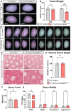
Impaired Sertoli cell proliferation in Nr5a1-Cre;Nedd4flox/flox mice. Testes (A) and Testis Weight (B, n = 14) of postnatal day (P) 60 Cre-negative littermate control (left, gray) and Nr5a1-Cre;Nedd4flox/flox (right, blue) mice. Hematoxylin and Eosin staining of Cre-negative (C) and Nr5a1-Cre;Nedd4flox/flox (D) P60 testes. Section immunofluorescence on P60 Cre-negative control (E) and Nr5a1-Cre;Nedd4flox/flox (F) testes stained for Sertoli cell marker SOX9 (green), granulosa cell marker FOXL2 (magenta). Scale bars = 100 μm. (G) Testis weights of control (gray) and Nr5a1-Cre;Nedd4flox/flox mice (blue) at P7 (n = 5), P14 (n = 6 control, 5 mutant), and P28 (n = 11 control, 9 mutant). Testis weights are normalized to body weight and are shown as a percentage of controls. (H) Section immunofluorescence on 15.5 dpc control and Nr5a1-Cre;Nedd4flox/flox testes stained for proliferation marker KI67 (green) and Sertoli cell marker SOX9 (magenta), counterstained with DAPI (gray). Scale bars = 100 μm. (I) Quantification of Sertoli cell proliferation of control (gray, n = 4) and Nr5a1-Cre;Nedd4flox/flox (blue, n = 5) fetal testes, shown as the proportion of proliferating Sertoli cells (KI67+ and SOX9+) within the total Sertoli cell pool (SOX9+). (J) Immunoblot on control (left) and Nr5a1-Cre;Nedd4flox/flox (Nedd4Δ/Δ, right) P60 testis lysates probed with NEDD4, PTEN, phosphorylated AKT (pAKT) and beta-actin (ACTB). Size markers (kilo Daltons) are shown on the lefthand side. (K) Quantification of pAKT immunoblot of control (gray, n = 3) and Nr5a1-Cre;Nedd4flox/flox (blue, n = 3) P60 testis lysates. Values are normalized to beta-actin and are shown relative to littermate testes. All graphs display mean ± SEM; t-test; *P < .05, ***P < .001, ****P < .0001.
Figure 4.
Figure 4.
Reduced testis size and abnormal testis morphology upon Sertoli cell–specific ablation of Nedd4. Testes (A) from control (left) and Sertoli cell–specific Nedd4 mutants (Amh-Cre;Nedd4flox/flox, right) at postnatal (P) day 15 (top) and P60 (bottom). (B) Testis weight of P15 and P60 Cre-negative littermate controls (left, gray, n = 9 [P15], n = 7 [P60]) and Amh-Cre;Nedd4flox/flox mice (right, red, n = 10 [P15], n = 11 [P60]) normalized to body weight and shown as a percentage of controls. (C) Testes of P60 controls (upper panel) and Amh-Cre;Nedd4flox/flox mice (lower panel) showing abnormal large blood vessels (red arrowhead) in mutants compared with their control littermates (white arrowhead). (D) Hematoxylin and eosin staining of P60 control and Amh-Cre;Nedd4flox/flox testes. Seminal vesicle weight (n = 5 control, 8 mutant) (E), sperm count (n = 6 control, 10 mutant) (F), and sperm motility measures (n = 6 control, 10 mutant) (G) of P60 control (left, gray) and Amh-Cre;Nedd4flox/flox mice (right, red). All graphs display mean ± SEM; t-test; ns = not significant, ***P < .001.
Figure 5.
Figure 5.
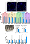
Perturbed adult Leydig cell differentiation in Nr5a1-Cre;Nedd4flox/flox testes. Section immunofluorescence on P60 Cre-negative control (A) and Nr5a1-Cre;Nedd4flox/flox (B) testes stained for Leydig cell markers CYP11A1 (green) and NR2F2 (magenta). Scale bars = 100 μm. (C) RT-qPCR analyses for fetal Leydig cell transcripts Ren1 and Crhr1 (red section), Leydig stem cell transcripts Nestin, Pdgfrb and Nr2f2 (yellow section), Leydig progenitor cell transcripts Akr1c14 and Srd5a1 (green section), immature adult Leydig cell transcript Hsd17b3 (light blue section), and adult Leydig cell transcripts Bhmt and Sult1e1 (purple section) on P60 control (gray, n = 10) and Nr5a1-Cre;Nedd4flox/flox testes (blue, n = 9). Anogenital distance (D, E, n = 7) and seminal vesicle weight (F, n = 11 control, 10 mutant) of P60 Cre-negative littermate control (Left, gray) and Nr5a1-Cre;Nedd4flox/flox mice (Right, blue). Seminal vesicle weight and anogenital distance are normalized to body weight and are shown as a percentage of controls. (G) RT-qPCR analyses for expression of genes encoding Ar, Lhr, Star, Cyp11a1, Cyp17a1, and Insl3 at P60 in Nr5a1-Cre;Nedd4flox/flox testes (blue, n = 5) and Cre-negative littermate controls (gray, n = 6). RT-qPCR values are normalized to Tbp and are expressed as fold change relative to controls. Mean ± SEM; t-test; ns = not significant, *P < .05, **P < .01, ****P < .0001.

References

    1. Windley SP, Wilhelm D. Signaling pathways involved in mammalian sex determination and gonad development. Sex Dev. 2015;9(6):297‐315. - PubMed
    1. Yao HH, Rodriguez KF. From Enrico Sertoli to freemartinism: the many phases of the master testis-determining celldagger. Biol Reprod. 2023;108(6):866‐870. - PMC - PubMed
    1. Yao HH, Whoriskey W, Capel B. Desert hedgehog/patched 1 signaling specifies fetal Leydig cell fate in testis organogenesis. Genes Dev. 2002;16(11):1433‐1440. - PMC - PubMed
    1. Brennan J, Tilmann C, Capel B. Pdgfr-alpha mediates testis cord organization and fetal Leydig cell development in the XY gonad. Genes Dev. 2003;17(6):800‐810. - PMC - PubMed
    1. Barsoum IB, Bingham NC, Parker KL, Jorgensen JS, Yao HH. Activation of the hedgehog pathway in the mouse fetal ovary leads to ectopic appearance of fetal Leydig cells and female pseudohermaphroditism. Dev Biol. 2009;329(1):96‐103. - PMC - PubMed

MeSH terms

Substances