

The role of PRP in the healing of disc degeneration and the effect of local anesthetics on PRP
- PMID: 40606908
- PMCID: PMC12213543
- DOI: 10.3389/fbioe.2025.1613148
The role of PRP in the healing of disc degeneration and the effect of local anesthetics on PRP
Abstract
Aim: This study aimed to investigate the regenerative effects of PRP on an experimental rat model of disc degeneration using histological and biochemical parameters. Additionally, we evaluated whether ropivacaine, a local anesthetic commonly used in clinical practice, affects the efficacy of PRP.
Methods: Rats were randomly divided into five groups as control and treatment groups. Disc degeneration models were established using appropriate procedures. On the intervention day, PRP was prepared from whole blood collected from the rats. PRP, PRP + ropivacaine, or ropivacaine alone was administered at the appropriate doses and according to standardized protocols.
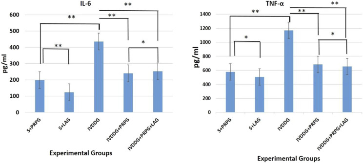
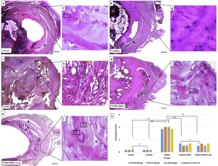
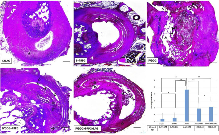
Results: In the untreated disc degeneration groups, annulus fibrosus (AF) and nucleus pulposus (NP) boundaries were indistinct, disc morphology was disrupted, collagen structures in the AF were degenerated or irregular, and vacuolization, interstitial edema, and necrotic tissue remnants were observed in the NP region. In contrast, in groups treated with PRP and PRP + ropivacaine, a reduction in edema and vacuolization, disappearance of necrotic tissue, restoration of distinct NP and AF boundaries, and decreased atrophy and cellular clustering in NP cells were observed. Biochemical analysis showed that IL-6 and TNF-α levels were within normal ranges in the groups treated with PRP and PRP + ropivacaine, whereas these levels remained elevated in the untreated disc degeneration groups, indicating ongoing effects of degeneration.
Conclusion: This study demonstrates the regenerative effects of PRP in disc degeneration through histological and biochemical parameters. Furthermore, the addition of ropivacaine to PRP did not exert any negative effects on PRP's regenerative properties.
Keywords: inflammation; intervertebral disc degeneration; local anesthesia; platelet-rich plasma; rat.
Copyright © 2025 Mert, Ikinci Keles, Aydin, Erol and Sonmez.
Conflict of interest statement
The authors declare that the research was conducted in the absence of any commercial or financial relationships that could be construed as a potential conflict of interest.
Figures
Similar articles
-
Suspension bioprinted whole intervertebral disc analogues enable regional stiffness- and hypoxia-regulated matrix secretion by primary human nucleus pulposus and annulus fibrosus cells.Acta Biomater. 2025 Jun 15;200:378-389. doi: 10.1016/j.actbio.2025.05.015. Epub 2025 May 7. Acta Biomater. 2025. PMID: 40339969
-
Risk factors for progression of nucleus pulposus degeneration in the lumbar intervertebral disc: a retrospective analysis using the disc signal intensity index.Spine J. 2025 Jul;25(7):1466-1473. doi: 10.1016/j.spinee.2025.01.036. Epub 2025 Feb 1. Spine J. 2025. PMID: 39900250
-
Ultrasound guidance for intervertebral disc location and paravertebral tissue swelling quantification in a mouse model of intervertebral disc herniation.Eur Spine J. 2025 Jun;34(6):2335-2346. doi: 10.1007/s00586-025-08843-8. Epub 2025 Apr 24. Eur Spine J. 2025. PMID: 40272495
-
Arthroplasty versus fusion in single-level cervical degenerative disc disease.Cochrane Database Syst Rev. 2012 Sep 12;(9):CD009173. doi: 10.1002/14651858.CD009173.pub2. Cochrane Database Syst Rev. 2012. Update in: Cochrane Database Syst Rev. 2015 May 21;(5):CD009173. doi: 10.1002/14651858.CD009173.pub3. PMID: 22972137 Updated.
-
Drugs for preventing postoperative nausea and vomiting in adults after general anaesthesia: a network meta-analysis.Cochrane Database Syst Rev. 2020 Oct 19;10(10):CD012859. doi: 10.1002/14651858.CD012859.pub2. Cochrane Database Syst Rev. 2020. PMID: 33075160 Free PMC article.
References
-
- Boncroft J. D. (1996). Theory and practice of histological techniques. Fourth Edition, 316. Tokyo.
-
- Browner B. D., Jupiter J. B., Krettek C., Anderson P. A. (2014). Sekeltal trauma; Basic science, management, and reconstruction. Saunders. EdsISBN 9780323294980.
LinkOut - more resources
Full Text Sources
Research Materials
Miscellaneous