

Short-term digital ocular massage may weaken corneal biomechanics
- PMID: 40606911
- PMCID: PMC12213799
- DOI: 10.3389/fbioe.2025.1582973
Short-term digital ocular massage may weaken corneal biomechanics
Abstract
Purpose: Digital ocular massage has been demonstrated to reduce intraocular pressure (IOP). However, its influence on corneal biomechanics remains unclear. In this study, a device employing Corneal Visualization Scheimpflug Technology (Corvis ST) was used to monitor changes in IOP and corneal biomechanics following short-term digital ocular massage in low and high myopes.
Methods: In total, 29 low myopes and 29 high myopes participated in this study. The right eyes (treatment eyes) underwent digital ocular massage for 5 min, whereas the left eyes (control eyes) remained closed during the procedure. Biomechanically-corrected IOP (bIOP) was measured in both eyes by using Corvis ST at three time points: before the ocular massage, immediately after the ocular massage, and 15 min post-massage. Dynamic corneal response (DCR) parameters were also monitored, namely, peak distance (PeakDist), highest concavity time (HCT), deformation amplitude (DA), deflection amplitude (DefleA), stress-strain index (SSI), time taken to reach the second applanation (A2T), and velocity required to reach the second applanation (A2V).
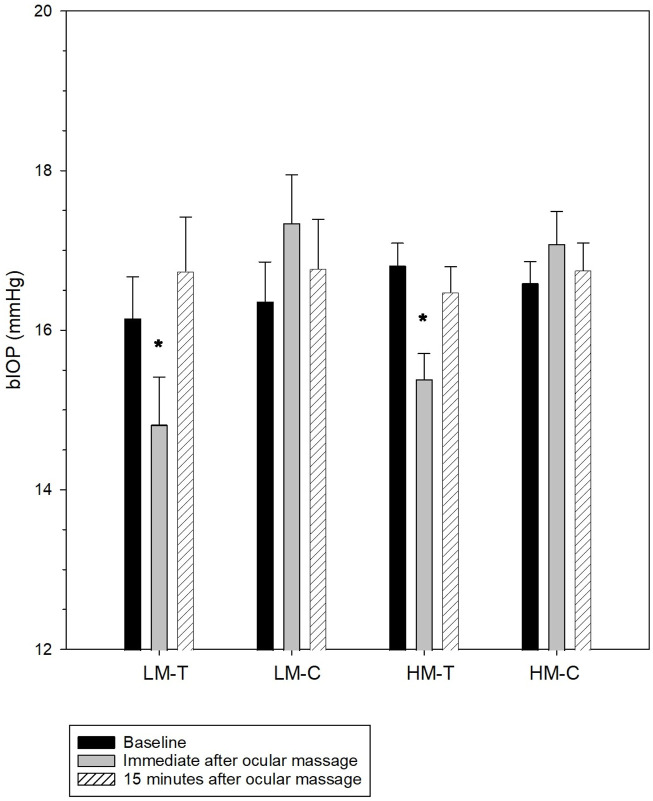
Results: At baseline, the participants exhibited comparable bIOP in both eyes. A significant reduction in bIOP was observed in the treatment eyes immediately after ocular massage (low myopes: 16.15 ± 2.79 vs. 14.82 ± 3.20 mmHg, p < 0.05; high myopes: 16.81 ± 1.51 vs. 15.39 ± 1.70 mmHg, p < 0.05). Corneal biomechanics at baseline were comparable between the treatment and control eyes. High myopes exhibited more deformable corneas, characterized by a shorter HCT (treatment eyes: 17.30 ± 0.41 vs. 17.72 ± 0.30 msec, p < 0.001; control eyes: 17.33 ± 0.32 vs. 17.55 ± 0.44 msec, p = 0.023), and lower SSI (treatment eyes: 0.739 ± 0.100 vs. 0.848 ± 0.114, p < 0.001; control eyes: 0.741 ± 0.103 vs. 0.858 ± 0.112, p < 0.001) than low myopes at baseline. Immediately after ocular massage, the treatment eyes in both groups exhibited shorter A2T, higher A2V, larger PeakDist, and higher DA and DefleA. Corneal biomechanics in the control eyes remained stable throughout. All DCR parameters returned to baseline levels 15 min after the ocular massage.
Conclusion: Short-term digital ocular massage results in a temporary reduction in bIOP. The observed changes, including shorter A2T, higher A2V, larger PeakDist, and greater DA and DefleA indicated a greater corneal deformability after ocular massage. These findings support the potential association between eye rubbing and the etiology or progression of keratoconus.
Keywords: Corneal biomechanics; Myopia; intraocular pressure; massage; stress strain index.
Copyright © 2025 Lam, Lee, Mui and Ng.
Conflict of interest statement
The authors declare that the research was conducted in the absence of any commercial or financial relationships that could be construed as a potential conflict of interest.
Figures
Similar articles
-
Comparison of intraocular pressure measurements obtained by Goldmann applanation tonometer, corvis ST and a conventional non-contact airpuff tonometer in eyes with previous myopic refractive surgery and correlation with corneal biomechanical parameters.Int Ophthalmol. 2025 Jun 6;45(1):232. doi: 10.1007/s10792-025-03598-z. Int Ophthalmol. 2025. PMID: 40478442
-
Corneal Biomechanical Characteristics in Myopes and Emmetropes Measured by Corvis ST: A Meta-Analysis.Am J Ophthalmol. 2024 Aug;264:154-161. doi: 10.1016/j.ajo.2024.03.024. Epub 2024 Mar 29. Am J Ophthalmol. 2024. PMID: 38556185
-
Safety and efficacy of stromal lenticule addition keratoplasty in the treatment of hyperopia: a retrospective study.BMC Ophthalmol. 2025 Jul 9;25(1):400. doi: 10.1186/s12886-025-04224-3. BMC Ophthalmol. 2025. PMID: 40634886 Free PMC article.
-
Intraocular Pressure Before and After Corneal Refractive Surgery: A Prospective Comparison of Corvis ST and Ocular Response Analyzer.J Glaucoma. 2024 Oct 1;33(10):780-784. doi: 10.1097/IJG.0000000000002434. Epub 2024 May 21. J Glaucoma. 2024. PMID: 38767500
-
Perioperative medications for preventing temporarily increased intraocular pressure after laser trabeculoplasty.Cochrane Database Syst Rev. 2017 Feb 23;2(2):CD010746. doi: 10.1002/14651858.CD010746.pub2. Cochrane Database Syst Rev. 2017. PMID: 28231380 Free PMC article.
References
LinkOut - more resources
Full Text Sources