

Case Report: Giant mesenteric hemolymphangioma enveloping the ileum
- PMID: 40606978
- PMCID: PMC12213793
- DOI: 10.3389/fonc.2025.1557916
Case Report: Giant mesenteric hemolymphangioma enveloping the ileum
Abstract
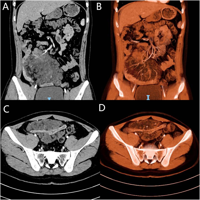
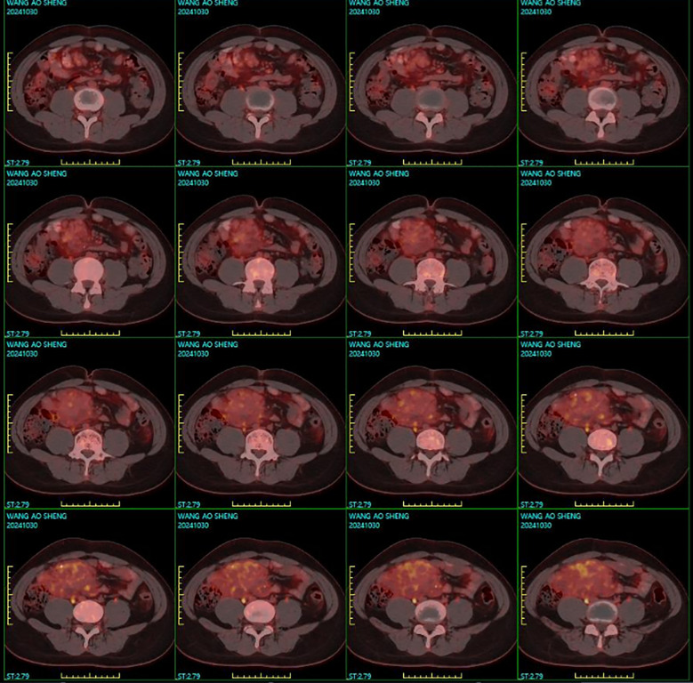
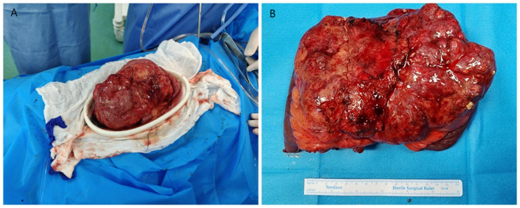
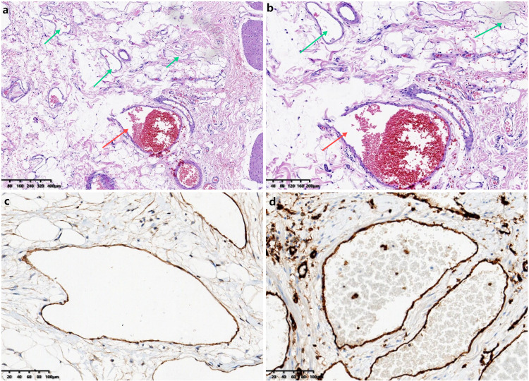
Hemolymphangiomas are rare, benign tumors arising from lymphatic and vascular tissues, most commonly found in subcutaneous and soft tissues, with their occurrence in the gastrointestinal tract, especially the mesentery, being uncommon. Due to their heterogeneous imaging features, these tumors are often misdiagnosed as malignancies, particularly when located in the abdominal cavity. We report a case of a 16-year-old male presenting with abdominal pain, vomiting, and diarrhea for 7 days. Imaging revealed a large, heterogeneous mass in the right lower abdomen, initially suspected to be a malignant mesenteric tumor. An ultrasound-guided biopsy and immunohistochemistry confirmed the diagnosis of hemolymphangioma. The patient underwent a combined laparoscopic and open surgical approach, including en bloc resection of the tumor, along with a segment of the ileum and surrounding mesentery. Histopathological examination verified the presence of lymphatic and vascular components consistent with hemolymphangioma. The patient recovered uneventfully and showed no recurrence at a 3-month follow-up. Hemolymphangiomas, although rare, should be considered in the differential diagnosis of mesenteric tumors.
Keywords: abdominal mass; case report; gastrointestinal involvement; hemolymphangioma; laparoscopic surgery.
Copyright © 2025 Yu, Liu, Yan, Shi, Yang, Wang, Li, Guo, Jiao, Zhang, Shi and Zhang.
Conflict of interest statement
The authors declare that the research was conducted in the absence of any commercial or financial relationships that could be construed as a potential conflict of interest.
Figures
Similar articles
-
Dietary interventions for recurrent abdominal pain in childhood.Cochrane Database Syst Rev. 2017 Mar 23;3(3):CD010972. doi: 10.1002/14651858.CD010972.pub2. Cochrane Database Syst Rev. 2017. PMID: 28334433 Free PMC article.
-
Does Augmenting Irradiated Autografts With Free Vascularized Fibula Graft in Patients With Bone Loss From a Malignant Tumor Achieve Union, Function, and Complication Rate Comparably to Patients Without Bone Loss and Augmentation When Reconstructing Intercalary Resections in the Lower Extremity?Clin Orthop Relat Res. 2025 Jun 26. doi: 10.1097/CORR.0000000000003599. Online ahead of print. Clin Orthop Relat Res. 2025. PMID: 40569278
-
Home treatment for mental health problems: a systematic review.Health Technol Assess. 2001;5(15):1-139. doi: 10.3310/hta5150. Health Technol Assess. 2001. PMID: 11532236
-
Ultrasonography for diagnosis of alcoholic cirrhosis in people with alcoholic liver disease.Cochrane Database Syst Rev. 2016 Mar 2;3(3):CD011602. doi: 10.1002/14651858.CD011602.pub2. Cochrane Database Syst Rev. 2016. PMID: 26934429 Free PMC article.
-
Drugs for preventing postoperative nausea and vomiting in adults after general anaesthesia: a network meta-analysis.Cochrane Database Syst Rev. 2020 Oct 19;10(10):CD012859. doi: 10.1002/14651858.CD012859.pub2. Cochrane Database Syst Rev. 2020. PMID: 33075160 Free PMC article.
References
Publication types
LinkOut - more resources
Full Text Sources
Research Materials