

Hemodynamic changes and their relationship with white matter hyperintensities in CSVD patients with cognitive impairment: a 4D flow study
- PMID: 40607188
- PMCID: PMC12213705
- DOI: 10.3389/fnagi.2025.1578288
Hemodynamic changes and their relationship with white matter hyperintensities in CSVD patients with cognitive impairment: a 4D flow study
Abstract
Objective: To observe the hemodynamics of intracranial arteries and veins in patients with cerebral small vessel disease (CSVD) with cognitive impairment (CI), and to explore the association between these flow features and white matter hyperintensities (WMH).
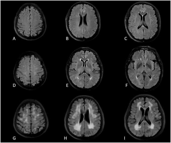
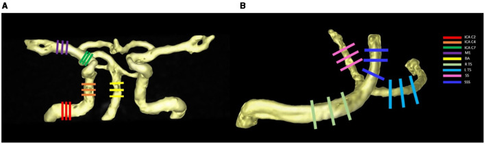
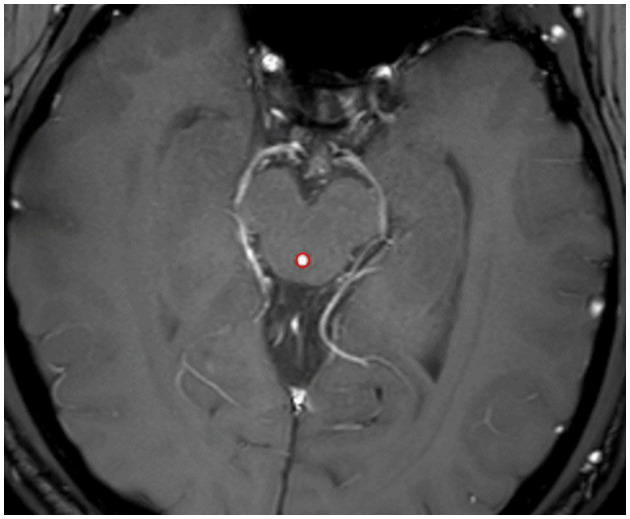
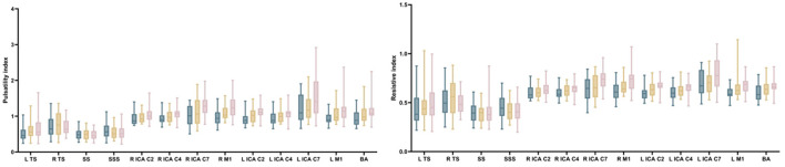
Materials and methods: A total of 53 patients with CSVD were included in the study, comprising 30 patients with CI (CI group) and 23 patients with non-CI (NCI group); Meanwhile, 25 age-matched cognitively healthy volunteers were recruited. WMH burden was evaluated using a 2D axial T2-FLAIR sequence. A 4D flow MRI was employed to measure intracranial hemodynamic features, including cross-sectional area, flow rate, blood flow velocity, wall shear stress (WSS), pulsatility index, and resistive index in the internal carotid artery (ICA), middle cerebral artery, basilar artery (BA), transverse sinus (TS), straight sinus (SS), and superior sagittal sinus (SSS). CSF-Q flow, a 2D PC MRI sequence, was performed to calculate the CSF fluid dynamics in the midbrain aqueduct.
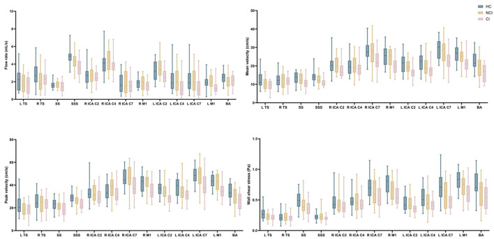
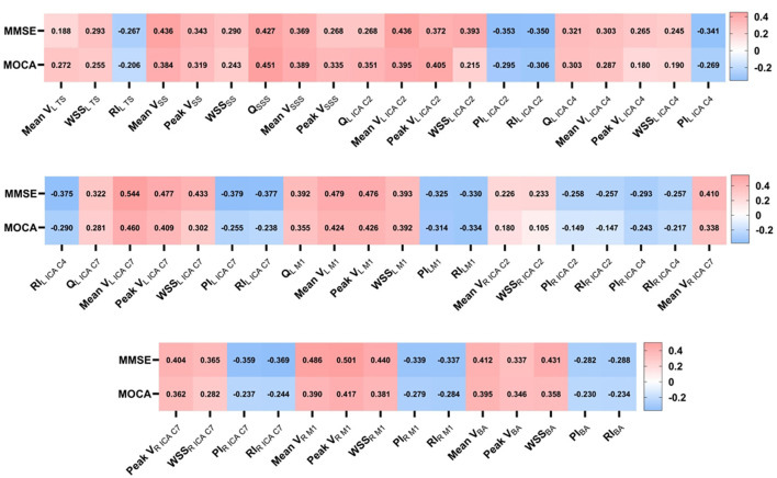
Results: The CSVD with CI population reported a statistically significant decrease in flow rate, blood flow velocity, and WSS, as well as an increase in PI, RI, CSF flow quantity, and velocity compared to age-matched cognitively healthy control participants. There was a moderately positive correlation between MMSE, MoCA score and flow rate, flow velocity, and WSS (r = 0.226-0.544, all P < 0.05), and a moderately negative correlation between MMSE, MoCA score and PI, RI (r = -0.230 to -0.406, all P < 0.05). Multiple linear regression indicated that, the flow rate and mean velocity in venous sinuses (β = -0.472 to -0.381, all P < 0.05) and the WSS in arterial segments (β = -0.771 to -0.441, all P < 0.05) had independently negative association with WMH burden; Meanwhile, a significant positive relationship was found between PI in arterial segments and specific-distributed WMH (PVWMH and S-CC WMH) (β = 0.239 to 0.356, all P < 0.05).
Conclusion: The intracranial hemodynamics were associated with CI and WMH in patients with CSVD. 4D flow MRI can be used as a non-invasive method to assess cerebrovascular hemodynamics and helps to identify patients who may benefit from interventions to improve the functions of the cerebral circulatory system and provides a potential new path for clinical treatment.
Keywords: 4D flow MRI; cerebral small vessel disease; cognitive impairment; hemodynamics; white matter hyperintensities.
Copyright © 2025 Cao, Yuan, Zhang, Zhao, Zhang, Chang, Song, Zhang, Hu and Miao.
Conflict of interest statement
The authors declare that the research was conducted in the absence of any commercial or financial relationships that could be construed as a potential conflict of interest.
Figures
Similar articles
-
Association of Mean Upper Cervical Spinal Cord Cross-Sectional Area With Cerebral Small Vessel Disease: A Community-Based Cohort Study.Stroke. 2024 Mar;55(3):687-695. doi: 10.1161/STROKEAHA.123.044666. Epub 2024 Jan 25. Stroke. 2024. PMID: 38269540
-
Hemodynamic analysis of non-stenotic middle cerebral artery in patients with cerebral ischemia based on 4D flow MRI.Front Neurosci. 2025 Jul 10;19:1502987. doi: 10.3389/fnins.2025.1502987. eCollection 2025. Front Neurosci. 2025. PMID: 40708689 Free PMC article.
-
Antithrombotic therapy to prevent cognitive decline in people with small vessel disease on neuroimaging but without dementia.Cochrane Database Syst Rev. 2022 Jul 14;7(7):CD012269. doi: 10.1002/14651858.CD012269.pub2. Cochrane Database Syst Rev. 2022. PMID: 35833913 Free PMC article.
-
MRI signatures associated with active ischemia and disease severity in cerebral small vessel disease.Neuroimage Rep. 2025 Aug 4;5(3):100281. doi: 10.1016/j.ynirp.2025.100281. eCollection 2025 Sep. Neuroimage Rep. 2025. PMID: 40809336 Free PMC article.
-
Systemic pharmacological treatments for chronic plaque psoriasis: a network meta-analysis.Cochrane Database Syst Rev. 2017 Dec 22;12(12):CD011535. doi: 10.1002/14651858.CD011535.pub2. Cochrane Database Syst Rev. 2017. Update in: Cochrane Database Syst Rev. 2020 Jan 9;1:CD011535. doi: 10.1002/14651858.CD011535.pub3. PMID: 29271481 Free PMC article. Updated.
References
-
- Alber J., Alladi S., Bae H. J., Barton D. A., Beckett L. A., Bell J. M., et al. (2019). White matter hyperintensities in vascular contributions to cognitive impairment and dementia (VCID): Knowledge gaps and opportunities. Alzheimers Dement (N Y). 5, 107–117. 10.1016/j.trci.2019.02.001 - DOI - PMC - PubMed
LinkOut - more resources
Full Text Sources
Research Materials
Miscellaneous