Review
. 2025 Jun 16.
doi: 10.1039/d5md00320b. Online ahead of print.

Small-molecule probes for imaging and impairing the Golgi apparatus in cancer

Affiliations
Review

Small-molecule probes for imaging and impairing the Golgi apparatus in cancer

Phanindra Kumar et al. RSC Med Chem. .

Abstract

The Golgi apparatus (GA) is one of the most important subcellular organelles controlling protein processing, post-translational modification and secretion. Dysregulation of the GA structure and function leads to multiple pathological states, including cancer development and metastasis. Consequently, visualizing GA dynamic structures and their impairment in cancer has emerged as a novel strategy for next-generation unorthodox cancer therapeutics. However, the major challenge in GA-mediated theranostic probe development is the specific targeting of the GA within the subcellular milieu due to the lack of GA-recognizing chemical entities. In this review, we delineated various chemical functionalities that are extensively used as GA-homing moieties. Moreover, we outlined GA imaging probes consisting of classical fluorophores as well as novel aggregation-induced emissive (AIE) probes tagged with GA-homing moieties. Furthermore, we described GA-impairing molecules that can damage GA morphology through chemotherapeutic and photodynamic therapy (PDT) in cancer. Finally, we addressed the current challenges in this emerging and underexplored field of GA-targeted theranostics and proposed potential solutions to guide future cancer therapeutics.


Conflict of interest statement

There are no conflicts to declare.

Figures

Fig. 1
Fig. 1. GA targeting chemical functionalities.
Fig. 2
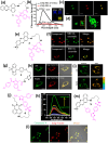
Fig. 2. (a) Chemical structure of the indomethacin-tagged fluorophore. (b) Fluorescence emission spectra of 1 (2.0 μM) in the absence and presence of COX-2. (c) Fluorescence images of 1 (5.0 μM) (green) and BODIPY TR C5-ceramide (5.0 μM) (red) in HeLa cells. (d) Changes in GA morphology of cancer cells during apoptosis, stained with compound 1 in MCF-7 cells. (e) Chemical structure of nile blue-tagged indomethacin probe (2). (f) Confocal images of MCF-7 and HeLa cells stained with compound 2 (green) and NBD C6-ceramide (red). (g) Chemical structure of the BODIPY-tagged indomethacin-based viscosity rotor in GA (3). (h) Confocal fluorescence images of compound 3 (green) and GA tracker red in LO2 and HepG2 cells. (i) Confocal fluorescence imaging and FLIM imaging for LO2 and HepG2 cells incubated with compound 3 for 30 min. (j) Chemical structure of napthalimide–celecoxib-based GA imaging probe (4). (k) Fluorescence emission spectra of compound 4 in the presence of COX-2. (l) Confocal images of compound 4 (green) with commercial BODIPY FL-C5-ceramide (red) in MCF-7. (m) Chemical structure of celecoxib–nile blue conjugated probe (5) for GA imaging. Fig. 2b–d have been reproduced with permission from ref. . Copyright 2018 American Chemical Society. Fig. 2f has been reproduced with permission from ref. . Copyright 2015, The Royal Society of Chemistry. Fig. 2h and i have been reproduced with permission from ref. . Copyright 2022, The Royal Society of Chemistry. Fig. 2k and l have been reproduced with permission from ref. . Copyright 2018 American Chemical Society.
Fig. 3
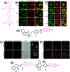
Fig. 3. (a) Chemical structures of 2-trifluoromethyl-7-aminoquinoline fluorophores (6 and 6′) for GA imaging. (b) Confocal fluorescence micrographs showing GA stained by 6 (green), BODIPY TR ceramide (red), and the merged images in different cancer cells. (c) Subcellular localization in synchronously dividing HeLa cells of 6 (green), BODIPY TR ceramide (red) and the merged images. (d) Chemical structure of ratiometric GA polarity detection probe. (e) Confocal ratiometric fluorescence imaging of compound 7 in HL-7702 cells without or with monensin stimulation. (f) TP fluorescence imaging of kidney tissues in control mice and in LPS-induced AKI mice. (g and h) Chemical structures of 2,6-dihydroxybenzoyl–hydrazone-based (8) and BODIPY-based fluorescence probe (9) for imaging GA in cancer cells. Fig. 3b and c have been reproduced with permission from ref. . Copyright 2019 American Chemical Society. Fig. 3e and f have been reproduced with permission from ref. . Copyright 2021. The Royal Society of Chemistry.
Fig. 4
Fig. 4. (a) Chemical structures of coumarin and napthalimide-based-lipid conjugated probes (10–12) for GA imaging. (b) Chemical structure of merocyanine–benzoyl difluoroboronate NIR probe (13) conjugated with cysteine for GA imaging. (c) Confocal fluorescence images of HL-7702 and SMMC-7721 cells stained with compound 13 (red). (d) Chemical structure of malononitrile-based GA imaging probe. (e) Confocal fluorescence images of compound 14 (red) in SH-SY5Y and LO2 cells stained with the GA tracker (green). Fig. 4c has been reproduced with permission from ref. . Copyright 2019 American Chemical Society. Fig. 4e has been reproduced with permission from ref. . Copyright 2024, Nature Publications.
Fig. 5
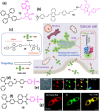
Fig. 5. (a) Chemical structure of the TPE-2′,3′-O-isopropylideneadenosine (Ade)-based AIEgen (15) for GA imaging. (b) Chemical structure of 9,10-distyrylanthracene–indomethacin-based AIEgen (16) (c) discrimination of COX-2 enzyme via specific compound 16 probe. (d) Chemical structure of phenylsulfonamide-based AIEgen (17) for GA imaging. (e) Confocal images of HeLa cells stained with compound 17 (green) and GA tracker (red). (f) Chemical structure of triphenylamine–naphthalimide-based “on–off–on” AIEgen (18) for GA imaging. (g) Confocal laser scanning microscopy images of HCT-116 cells incubated with compound 18 (green) for 30 min, followed by staining GA with GA-tracker red dye. Fig. 5c has been reproduced with permission from ref. . Copyright 2022, Chinese Chemical Society. Fig. 5e has been reproduced with permission from ref. . Copyright 2021, Royal Society of Chemistry. Fig. 5g has been reproduced with permission from ref. . Copyright 2025, American Chemical Society.
Fig. 6
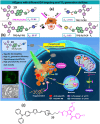
Fig. 6. (a) Chemical structures of cyanovinyl–methylpyridinium-based photosensitizer AIEgens for GA targeting. (b) Schematic of AIEgen-induced GA stress and the crosstalk between GA and mitochondria during cell apoptosis upon PDT. (c) Chemical structure of AIEgen-based photosensitizer containing indomethacin and triphenylamine–thiophene–vinyl–pyridinium cation. Fig. 6a and b have been reproduced with permission from ref. . Copyright 2022, Nature Publications.
Fig. 7
Fig. 7. (a and b) Chemical structures of phospho and thioacetate peptides containing nitrobenzoxadiazole (NBD) for targeting GA. (c) Confocal images of the HeLa cells having peptide 22 homing into the GA. (d) Chemical structure of zinc phthalocyanines–indomethacin conjugate photosensitizer for GA targeting. Fig. 7c has been reproduced with permission from ref. . Copyright 2022, American Chemical Society.
None
Phanindra Kumar
None
Poulomi Sengupta
None
Sudipta Basu

Similar articles

References

    1. Baker J. R. Nature. 1951;168:1089–1090. - PubMed
    1. Featherstone C. Science. 1998;282:2172–2174. - PubMed
    1. Li J. Ahat E. Wang Y. Results Probl. Cell Differ. 2019;67:441–485. - PMC - PubMed
    1. Zappa F. Failli M. De Matteis M. A. Curr. Opin. Cell Biol. 2018;50:102–116. - PubMed
    1. Eisenberg-Lerner A. Benyair A. Hizkiahou S. Nudel T. N. Maor-Nof H. Kramer T. Yayon N. Ulman Y. Yarden E. G. Elazar Y. Nat. Commun. 2020;11:409. - PMC - PubMed

LinkOut - more resources