Review
. 2025 Jun 18:16:1609509.
doi: 10.3389/fimmu.2025.1609509. eCollection 2025.

Revisiting pulmonary fibrosis: inflammatory dynamics of the lipofibroblast-to-inflammatory lipofibroblast-to-activated myofibroblast reversible switch

Affiliations
Review

Revisiting pulmonary fibrosis: inflammatory dynamics of the lipofibroblast-to-inflammatory lipofibroblast-to-activated myofibroblast reversible switch

Georgios-Dimitrios Panagiotidis et al. Front Immunol. .

Abstract

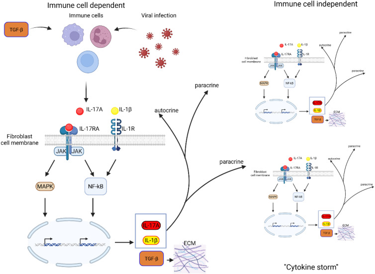
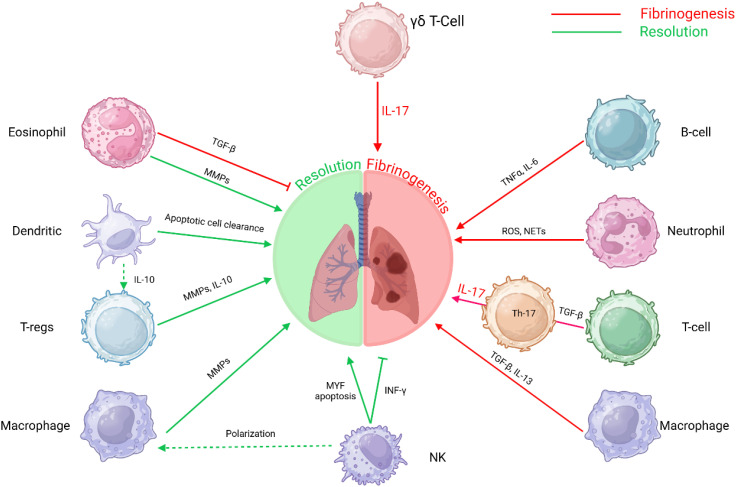
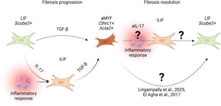
Idiopathic pulmonary fibrosis (IPF) is a chronic, progressive interstitial lung disease characterized by excessive extracellular matrix (ECM) deposition and irreversible lung damage. A key driver of disease progression is the phenotypic shift of lipofibroblasts (LIFs) into activated myofibroblasts (aMYFs), triggered by sustained epithelial injury, caused by inflammation, oxidative stress, viral infections (e.g., influenza, SARS-CoV-2), and metabolic dysfunction. Emerging evidence demonstrates that this transition is reversible, with pharmacological agents that promote aMYF-to-LIF reprogramming contributing to fibrosis resolution. The identification of inflammatory lipofibroblasts (iLIFs) highlights the importance of inflammation in fibrosis progression. Inflammation, mediated by IL-1β, IL-17A, and TGF- β, sustain aMYF activation, while immune cells shape fibrosis formation. This review combines current insights on the cellular and molecular pathways controlling fibroblast differentiation, highlighting key metabolic, immunologic, and oxidative stress-modulating targets for therapeutic intervention. Understanding and manipulating the LIF-iLIF-aMYF axis offers a promising strategy for reversing fibrosis and restoring pulmonary homeostasis in IPF.

Keywords: IL-17A; TGF-β; activated myofibroblast; idiopathic pulmonary fibrosis; inflammation; inflammatory lipofibroblast; lipofibroblast; virus infection.


Conflict of interest statement

The authors declare that the research was conducted in the absence of any commercial or financial relationships that could be construed as a potential conflict of interest.

Figures

Figure 1
Figure 1
Schematic representation of lung fibrosis progression and its cellular and metabolic mechanisms. In healthy lung, alveolar epithelial cells (AT1 and AT2) maintain normal gas exchange, with lipofibroblasts (LIF) providing support to the alveolar niche. Under persistent oxidative stress (ROS), inflammation, virus infection and metabolic dysregulation, LIF transition into intermediate inflammatory lipofibroblasts (iLIFs) (18), leading to impaired alveolar epithelial stem cell niche. Persistent injury, inflammation and activation of myofibroblasts (aMYF) result in fibrosis, disrupting alveolar architecture and leading to loss of gas exchange. The progressive pathological changes ultimately contribute to impaired lung function. EAT2/1: early intermediate AT2/AT1, LAT2/AT1: late intermediate AT2/AT1.
Figure 2
Figure 2
Schematic representation of IL-17A-TGF-β positive feedback loop in fibroblasts. TGF-β stimulation of immune cells as well as inflammatory response to virus infection induces IL-17A. IL-17A binds to its receptor IL-17RA, activating downstream signaling pathways, including MAPK and NF-κB, which drive gene transcription. This leads to the production of IL-17A, IL-1β, and TGF-β, amplifying inflammatory responses. IL-17A and IL-1β act in a paracrine and autocrine reinforcing the inflammatory loop. Increased TGF-β expression promotes ECM deposition, contributing to fibrosis and tissue remodeling. Persistent activation of these pathways may lead to excessive fibrotic responses, disrupting normal tissue architecture and function.
Figure 3
Figure 3
Immune cell interactions in fibrosis: This schematic depicts the intricate roles of various immune cells in the progression and resolution of fibrosis. The interplay between these immune cells and molecular pathways underscores the multifaceted nature of fibrosis, involving immune regulation, tissue remodeling, and inflammatory responses. T-helper 17 T-lymphocytes Th17 T-cells, B-lymphocytes B-cells, Gamma delta T-lymphocytes γδ T-Cells, T-regulatory cells (T-regs) and Natural Killer cells (NK).
Figure 4
Figure 4
Proposed model of fibrosis progression and resolution. During fibrosis progression, lung alveolar lipofibroblasts (LIF Scube2+ ) transition into an inflammatory LIF-expressing state (iLIF) under IL-17-mediated inflammation (Tsukui et al., 2024). iLIF, driven by TGF-β, differentiate into activated myofibroblasts (aMYF, Cthrc1+ Acta2+ ), promoting fibrosis. Resolution involves myofibroblast reversion to lipofibroblasts (LIF) (25, 26, 52). Tsukui et al., 2024 suggest IL-17 inhibition (aIL-17) modulates iLIF, yet mechanisms governing iLIF reversion to LIF remain unclear.

Similar articles

References

    1. Richeldi L, Collard HR, Jones MG. Idiopathic pulmonary fibrosis. Lancet. (2017) 389:1941–52. doi: 10.1016/S0140-6736(17)30866-8 - DOI - PubMed
    1. Wynn TA, Ramalingam TR. Mechanisms of fibrosis: Therapeutic translation for fibrotic disease. Nat Med. (2012) 18:1028–40. doi: 10.1038/nm.2807 - DOI - PMC - PubMed
    1. Moss BJ, Ryter SW, Rosas IO. Pathogenic mechanisms underlying idiopathic pulmonary fibrosis. Annu Rev Pathol. (2022) 17:515–46. doi: 10.1146/annurev-pathol-042320-030240 - DOI - PubMed
    1. Somogyi V, Chaudhuri N, Torrisi SE, Kahn N, Müller V, Kreuter M. The therapy of idiopathic pulmonary fibrosis: What is next? Eur Respir Rev. (2019) 28. doi: 10.1183/16000617.0021-2019 - DOI - PMC - PubMed
    1. Wight TN, Potter-Perigo S. The extracellular matrix: an active or passive player in fibrosis? Am J Physiol Gastrointest Liver Physiol. (2011) 301:G950–5. doi: 10.1152/ajpgi.00132.2011 - DOI - PMC - PubMed

LinkOut - more resources