. 2025 Jul 22;99(7):e0011025.
doi: 10.1128/jvi.00110-25. Epub 2025 Jul 3.

Birds of a feather flock together: structural characterization of red-crowned crane and turkey aveparvoviruses

Affiliations

Birds of a feather flock together: structural characterization of red-crowned crane and turkey aveparvoviruses

Jane Hsi et al. J Virol. .

Abstract

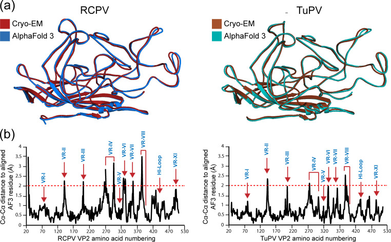
The parvoviruses have non-enveloped T = 1 icosahedral capsids that are ~25 nm in diameter, which package linear single-stranded DNA and infect a wide range of hosts. To date, parvoviruses affecting birds have been identified in two genera: Ave- and Dependoparvovirus, and no capsid structures have been determined for any member of Aveparvovirus. This study investigates two bird viruses of this genus: red-crowned crane parvovirus (RCPV) and turkey parvovirus (TuPV). While the pathogenicity of RCPV is currently unknown, TuPV has been associated with gastrointestinal diseases, especially in juvenile birds. High-resolution structures of the RCPV and TuPV capsids were determined by cryo-electron microscopy at a resolution of 2.66 Å and 2.35 Å, respectively. A structural comparison of the RCPV and TuPV capsids shows that they exhibit many conserved features, such as a channel at the five-fold symmetry axis and surface depressions at the two-fold axis, as previously observed in parvoviruses from other genera. However, major structural differences were observed at the three-fold axes, with both RCPV and TuPV displaying recessed protrusions. In addition, terminal sialic acid was identified as a potential glycan receptor for RCPV. This study extends the architectural portfolio of structural parvovirology and will provide insights into parvoviral diversity and characterization of these viruses.IMPORTANCEThis study presents the capsid structures of two aveparvoviruses, red-crowned crane parvovirus (RCPV) and turkey parvovirus (TuPV), extending the structural repertoire of the Parvoviridae. While the pathogenicity of RCPV is unknown, the red-crowned cranes are among the rarest crane species. To date, very few virological studies have been conducted for this rare avian species, and understanding their virome could contribute to conservation efforts. Additionally, several studies have previously suggested that TuPV is associated with cases of enteric disease syndrome. To date, no commercial antivirals or vaccines are available for TuPV. The structural characterization of its capsid may contribute toward the development of a treatment to control the spread of infection.

Keywords: Aveparvovirus; RCPV; TuPV; capsid; cryo-EM; parvovirus; pathogen; red-crowned crane; turkey.


Conflict of interest statement

The authors declare no conflict of interest .

Figures

Fig 1
Fig 1
Production/purification and structure determination of RCPV and TuPV. (a) Top panel: an SDS-PAGE of RCPV displaying a band at 60 kDa consistent with the size of VP2 (left side). A cryo-EM micrograph of the same purified sample exhibiting intact capsids that are ~25 nm in diameter. Bottom panel: the capsid surface density map of RCPV determined via cryo-EM reconstruction, which was contoured at a sigma (σ) threshold = 2. The resolution of the structure is calculated based on an FSC threshold = 0.143. The surface density map is radially colored (blue to red) based on the distance to the center of the particle, as shown in the scale bar below. The icosahedral two-, three-, and five-fold symmetry axes are indicated on the RCPV capsid map. (b) Depiction as in panel (a) for TuPV.
Fig 2
Fig 2
The VP2 monomers of RCPV and TuPV. (a) Seven amino acid residues modeled for β strand in VR-II inside their respective density maps at a sigma (σ) threshold of 2.5 (in black mesh). (b) Seven amino acid residues modeled for VR-IX surface loop inside their respective density maps at a sigma (σ) threshold of 2.5 (in black mesh). All amino acid residues in this figure are labeled and shown as stick representations and colored according to the following color scheme by atom types: C = yellow, O = red, and N = blue. The images were generated utilizing UCSF-Chimera. (c) The VP2 monomer structures are depicted as ribbon diagrams shown inside semi-transparent surface representations. Secondary structure elements, the N- and C-termini, and VRs are labeled. (d) Location of the VRs on the surface of the capsid. The colors are as indicated. These images were generated using PyMOL (41).
Fig 3
Fig 3
Bivalent cation interaction sites on the RCPV and TuPV VLPs. The amino acid residues are labeled and shown as stick representations and colored according to the following color scheme based on atom types: C = yellow, O = red, and N = blue. Bivalent cation (Ca2+ or Mg2+) is shown as a sphere and colored in light green. Water molecules are represented as a sphere and colored in red. The images were generated with UCSF-Chimera.
Fig 4
Fig 4
RCPV is a sialic acid binder. Fluorescent-labeled VLPs were incubated with CHO Pro5 and Lec2 cell lines that display terminal sialic acid or galactose, respectively. The binding of the VLPs was determined via an FACS-based assay. Shown are example FACS histograms for control cells and cells incubated with RCPV and SAAV, and the percentages of bound cells are provided. Below is a summary of the triplicate experiments. The surface glycans of the cell lines are represented as symbols.
Fig 5
Fig 5
Comparison of AlphaFold 3 predictions against cryo-EM determined VP structures of RCPV and TuPV. (a) Structural superposition of RCPV (left) and TuPV (right) determined by cryo-EM and AlphaFold 3 is shown as ribbon diagrams. The images were generated in PyMOL (41). (b) The Cα-Cα distance plot for the RCPV (left) and TuPV (right) amino acid residues relative to the AlphaFold 3 predicted structures when the VP structures are superposed. The assigned variable regions are labeled in blue.
Fig 6
Fig 6
The Aveparvovirus genus. (a) A phylogenetic tree of all Aveparvovirus members was generated online (ngphylogeny.fr) utilizing the VP2 amino acid sequences as the input (62). Amino acid sequence identities were compared for the VP1 and VP2 of all aveparvoviruses vs RCPV. (b) A comparison of AlphaFold 3 generated surface representations of ChPV, PfPV, PiPV, and MAPV to RCPV and TuPV is shown. The superposition of the aveparvoviruses VP structures is depicted as cartoon ribbon diagrams below. The VRs, β-strands, and the icosahedral symmetry axes are labeled. (c) Table summarizing the number of amino acid residues for each of the VRs for all aveparvoviruses.
Fig 7
Fig 7
Comparison of RCPV VP to VPs from other genera in Parvovirinae. (a) The major VP monomer structures are depicted as ribbon diagrams for RCPV against a representative member of six other Parvovirinae genera. (b) Table of Cα distances of major VP and amino acid sequence identity of VP1 and major VP of RCPV compared to CPV, AMDV, HBoV1, QAAV, PARV4, and B19.

References

    1. Samantha JL, Florian D, Paul D. 2024. A Brief History of Bird Flu. Philosophical Transactions of the Royal Society B: Biological Sciences. Retrieved 1 Jun 2024. doi: 10.1098/rstb.2018.0257 - DOI
    1. Susan FC. 2023. ICTV virus taxonomy profile: parvoviridae. Microbiology Society. Retrieved 16 Feb 2023. doi: 10.1099/jgv.0.001212 - DOI
    1. Pénzes JJ, Söderlund-Venermo M, Canuti M, Eis-Hübinger AM, Hughes J, Cotmore SF, Harrach B. 2020. Reorganizing the family Parvoviridae: a revised taxonomy independent of the canonical approach based on host association. Arch Virol 165:2133–2146. doi: 10.1007/s00705-020-04632-4 - DOI - PubMed
    1. Kapgate SS, Kumanan K, Vijayarani K, Barbuddhe SB. 2018. Avian parvovirus: classification, phylogeny, pathogenesis and diagnosis. Avian Pathol 47:536–545. doi: 10.1080/03079457.2018.1517938 - DOI - PubMed
    1. Vibin J, Chamings A, Klaassen M, Bhatta TR, Alexandersen S. 2020. Metagenomic characterisation of avian parvoviruses and picornaviruses from Australian wild ducks. Sci Rep 10:12800. doi: 10.1038/s41598-020-69557-z - DOI - PMC - PubMed

Substances

LinkOut - more resources