. 2025 Aug 13;16(8):e0124925.
doi: 10.1128/mbio.01249-25. Epub 2025 Jul 3.

IFNγ-inducible Gbp4 and Irgb6 contribute to experimental cerebral malaria pathology in the olfactory bulb

Affiliations

IFNγ-inducible Gbp4 and Irgb6 contribute to experimental cerebral malaria pathology in the olfactory bulb

Julia Matsuo-Dapaah et al. mBio. .

Abstract

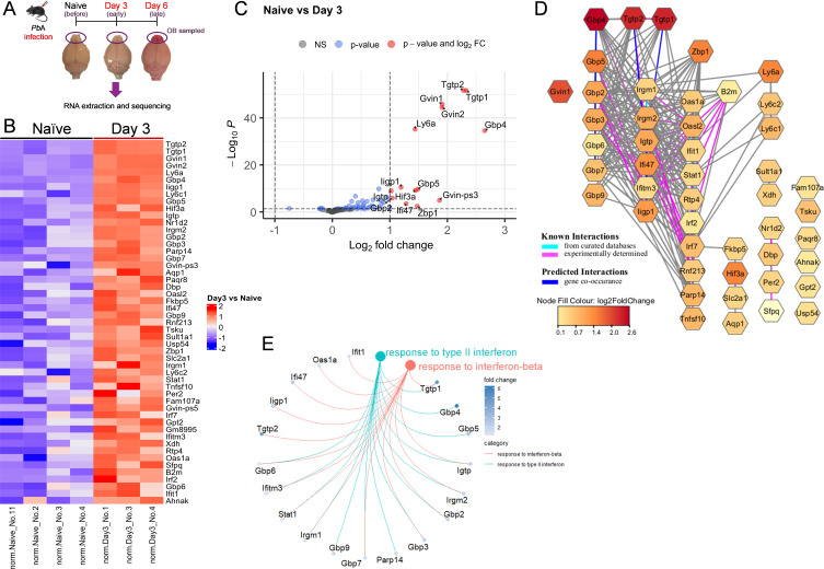
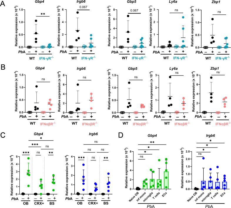
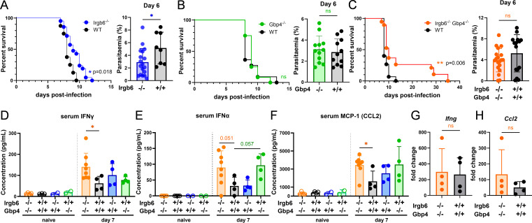
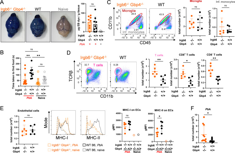
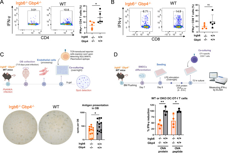
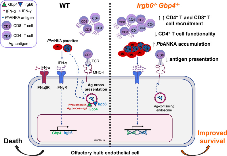
Cerebral malaria (CM) is a severe and often fatal complication of Plasmodium falciparum infection. Although much progress has been made in understanding CM, the precise pathogenesis remains elusive. The olfactory bulb (OB) has emerged as a critical site of immunopathology in experimental cerebral malaria (ECM) models, but its contribution to disease progression is not fully understood. To investigate the molecular mechanisms driving early ECM pathogenesis, we conducted transcriptomic profiling of the OB to identify key genes associated with disease onset. Our analysis revealed significant early upregulation of interferon (IFN)-inducible GTPases, particularly Irgb6 and Gbp4, effectors downstream of IFN-γ but not IFN-α/β signaling, suggesting their involvement in ECM pathology. Using Gbp4-/-, Irgb6-/-, and double knockout (Irgb6-/- Gbp4-/-) mice, we identified a pathological role for these GTPases. Mechanistically, we found that double-knockout mice exhibited increased infiltration of CD4+ and CD8+ T cells into the brain but with reduced T cell functionality and impaired antigen presentation by endothelial cells, leading to enhanced parasite accumulation in the OB. This disruption in immune regulation ultimately conferred improved survival in the Irgb6-/- Gbp4-/- mice and indicated the pathological impact of Gbp4 and Irgb6 in ECM. These findings reveal that Gbp4 and Irgb6 play important roles in the early immunopathogenesis of ECM by modulating antigen processing and presentation in the OB, thereby shaping immune cell dynamics. Our work shows the dual role of Irgb6 and Gbp4 GTPases in host defence and immunopathology and offers new insights into ECM mechanisms and antigen presentation.IMPORTANCECerebral malaria (CM) arises from an excessive inflammatory response and blood-brain-barrier (BBB) dysfunction in Plasmodium-infected hosts, but the precise mechanisms driving early-stage pathogenesis remain unclear. Through RNA sequencing of the olfactory bulb (OB) in a murine experimental cerebral malaria (ECM) model, we identified the early upregulation of interferon (IFN)-inducible GTPases, Irgb6 and Gbp4, key effectors downstream of IFN-γ signaling. Our results demonstrate that Gbp4 and Irgb6 synergistically contribute to ECM pathology by regulating antigen cross-presentation in endothelial cells. This dysregulation leads to abnormal parasite burden and alters the accumulation of CD4+ and CD8+ T cells in the brain via the OB, further perturbing inflammation. Our findings suggest a novel mechanism in CM and emphasize the pivotal roles of Gbp4 and Irgb6 in promoting cell-autonomous immune responses that, in turn, escalate pathological inflammation. Our study offers insights into how dysregulated immune responses drive CM progression and suggests potential therapeutic targets to mitigate fatal outcomes.

Keywords: GTPases; Gbp4; Irgb6; cerebral malaria; olfactory bulb.


Conflict of interest statement

The authors declare no conflict of interest.

Figures

Fig 1
Fig 1
Transcriptomic analysis of the olfactory bulb identifies Gbp4 and Irgb6 as interferon-inducible GTPases upregulated early during ECM. (A) Schematic outline of the experimental design. Olfactory bulbs were collected at different time points after PbA infection for RNA extraction, sequencing, and transcriptomic analysis (n = 3–4 mice/group on days 0, 3, and 6 post-infection). (B) Heatmap of differentially expressed genes in the OBs of uninfected versus PbA-infected mice on day 3 post-infection. Fifty-one genes were significantly upregulated, and five genes were downregulated on day 3 compared to uninfected samples (Padj < 0.05). (C) Volcano plot of differentially expressed genes in the OBs of uninfected versus PbA-infected mice on day 3 post-infection. Significantly upregulated genes (fold-change ≥ 1.0 and adjusted P-value < 0.05) are indicated on the upper right (red dots). Horizontal dotted line indicates the cut-off of adjusted P-values. Vertical dotted line indicates the cut-off of log2 fold-change (−1.0 to 1.0). (D) STRING association network of differentially expressed genes upregulated on day 3 post-PbA infection compared to naïve. Node fill color indicates log2 fold-change. Edges depict interactions, with aqua for known interactions from curated databases, lavender for experimentally determined known interactions, and blue for predicted gene co-occurrence. (E) Gene concept network of two gene ontology biological processes (GO:BP) terms “response to type II interferon” and “response to interferon-beta” enriched in genes differentially upregulated on day 3 post-PbA infection compared to naïve.
Fig 2
Fig 2
Gbp4 and Irgb6 are expressed in various OB-resident and OB-infiltrating cells in response to IFN-γ, but not type I IFNs. WT and IFN-γR−/− and IFNα/βR−/− mice were infected with PbA, and OB were analyzed on days 0 and 6 post-infection. (A and B) Relative expressions of early expressed genes Gbp4, Irgb6, Gbp5, Ly6a, and Zbp1 in OBs from WT and IFN-γR−/− mice (A), and IFNα/βR−/− mice on days 0 and 6 post-infection with PbA parasites. (C) Relative expressions of Gbp4 and Irgb6 in OB, cortex, and brainstem of WT mice at days 0 and 6 post-infection with PbA parasites. (D) RNAs were extracted from FACS-sorted infiltrating monocytes, T cells, microglia, and endothelial cells from the olfactory bulb of PbA-infected mice, and the relative expression of Gbp4 and Irgb6 was measured. Data are shown as mean ± SD from n = 4–5 mice/group (A through C). Data are pooled from four independent experiments and shown as mean ± SD (pooled from eight mice, collected day 6, moribund) (D). Mann-Whitney test (A, B), one-way ANOVA with multiple comparisons test (C), and Kruskal-Wallis test (D) were used for the statistical analysis. *, P < 0.05; **, P < 0.01; ***, P < 0.001; ns, not significant.
Fig 3
Fig 3
Gbp4 and Irgb6 contribute to ECM development. Groups of mice were infected with 105 PbA-GFP iRBCs. (A) Survival and day 6 parasitemia of Irgb6−/− (n = 21) and WT (n = 8) mice. (B) Survival and day 6 parasitemia of Gbp4−/− (n = 12) and WT (n = 11) mice. (C) Survival and day 6 parasitemia of Irgb6−/− Gbp4−/− (n = 19) and WT (n = 15) mice. (D through F) Detection of IFN-γ (D), IFN-α (E), and MCP-1/Ccl2 (F) protein levels in the sera of Irgb6−/−, Gbp4−/−, Irgb6−/− Gbp4−/−, and WT mice at days 0 and 7 post-infection (n = 4–8 mice/group) by either multiplex cytokine detection or enzyme-linked immunosorbent assay (ELISA). (G and H) Relative expressions of Ifng (G) and Ccl2 (H) in the olfactory bulb of WT and Irgb6−/− Gbp4−/− mice on day 6 post-infection with 106 PbA iRBCs relative to naïve (n = 4 mice/group). Survival comparisons between two groups were analyzed by the log-rank Mantel-Cox test (A through C). Data are shown as mean ± SD. Mann-Whitney test was used for statistical analysis. *, P < 0.05; **, P < 0.01; ns, not significant.
Fig 4
Fig 4
In vivo assessment of infected Irgb6−/− Gbp4−/− mice during ECM. Groups of Irgb6−/− Gbp4−/− and WT mice were infected for the in vivo assessment of ECM. (A) Representative images of Evans blue dye injected mice from naïve and infected groups and Evans blue dye quantification in OB of PbA-infected WT, Irgb6−/− Gbp4−/−, and naïve WT mice on day 8 post-infection (n = 4–8 mice/group). (B) Buried food test of naïve and PbA-infected WT and Irgb6−/− Gbp4−/− mice on day 7 post-infection. Time taken to find hidden food is shown in seconds (n = 9–13 mice/group). (C and D) Representative flow cytometry plots and absolute cell numbers of infiltrating monocytes and microglia (C), and TCRβ+, CD8+ T, and CD8 T cells (D) in the olfactory bulb of WT and Irgb6−/− Gbp4−/− mice at day 8 post-infection. Each data point represents a sample pooled from 2 to 3 mice, normalized by the number of mice used (n = 6 mice/group). (E) Absolute cell counts of endothelial cells and their MHC-I and -II expression are shown as geometric mean fluorescence intensity (gMFI), in the olfactory bulb of WT and Irgb6−/− Gbp4−/− mice at day 8 post-infection. (F) Quantified numbers of GFP-PbA in the olfactory bulb of WT and Irgb6−/− Gbp4−/− mice at day 8 post-infection. Each data point represents an OB randomly pooled from 2 to 3 mice in each group and normalized by the number of mice. Scatter plots present the mean ± SD. Data were analyzed by using one-way ANOVA with multiple comparisons test (B), Student’s t test (A, C through E), and Mann-Whitney test (F). *, P < 0.05; **, P < 0.01; ns, not significant.
Fig 5
Fig 5
Despite higher brain infiltrating CD4+ and CD8+ T cells, less cross-presentation results in less IFN-γ production in Irgb6−/− Gbp4−/− mice OBs. (A and B) Flow cytometry analysis of IFN-γ+ CD4 T cells (A) and IFN-γ+ CD8 T cells (B) in the olfactory bulb of WT and Irgb6−/− Gbp4−/− mice on day 8 after PbA infection. Each dot represents a sample pooled from 2 to 4 mice. Representative flow cytometry plots are shown. (C) Schematic diagram of the experimental protocol and the results of ex vivo endothelial cell antigen cross-presentation assay from PbA-infected WT and Irgb6−/− Gbp4−/− mice in the OB. Representative wells are shown. Data were pooled from three independent experiments, n = 12 for each group. Each dot represents a sample from one mouse. (D) Schematic diagram of the experimental protocol and IFN-γ results of BMDC: OT-I CD8 T cell co-culture assay. LPS-activated BMDCs from WT and Irgb6−/− Gbp4−/− mice were induced with OVA or OVA257–264 peptide prior to co-culture with purified OT-I CD8+ T cells for 3 days at a 1:1 ratio. Concentrations of IFN-γ in the culture supernatants were measured by ELISA. Data were depicted as %IFN-γ-reduction compared to WT mice values and pooled from the two independent experiments, n = 4 for each group. Each dot represents a sample from one mouse. Data are shown as mean ± SD and analyzed with Student’s t test. *, P < 0.05; **, P < 0.01; ns, not significant.
Fig 6
Fig 6
Proposed model for the role of GTPases Gbp4 and Irgb6 during experimental cerebral malaria in the OB. In WT mice, infection with PbA triggers systemic production of IFN-γ, which binds to IFN-γ receptors on endothelial cells in the OB and induces the expression of Gbp4 and Irgb6. These endothelial cells then process and cross-present PbA antigens to infiltrating T cells, leading to the ECM pathology. In Irgb6−/− Gbp4−/− mice, the absence of these GTPases results in increased PbA accumulation, increased CD4+ and CD8+ T cell infiltration, but impaired T cell functionality. Additionally, the disruption of PbA antigen cross-presentation in these double-knockout mice suggests that Gbp4 and Irgb6 play roles in antigen processing and presentation. These findings overall indicate that Gbp4 and Irgb6 are key components in regulating immune responses in the OB, while helping to coordinate effective pathogen control, which in turn leads to excessive immune activation and pathology.

Similar articles

References

    1. World Health Organization (WHO). 2024. Malaria Report 2024
    1. Idro R, Jenkins NE, Newton CRJC. 2005. Pathogenesis, clinical features, and neurological outcome of cerebral malaria. Lancet Neurol 4:827–840. doi: 10.1016/S1474-4422(05)70247-7 - DOI - PubMed
    1. Hadjilaou A, Brandi J, Riehn M, Friese MA, Jacobs T. 2023. Pathogenetic mechanisms and treatment targets in cerebral malaria. Nat Rev Neurol 19:688–709. doi: 10.1038/s41582-023-00881-4 - DOI - PubMed
    1. Coban C, Lee MSJ, Ishii KJ. 2018. Tissue-specific immunopathology during malaria infection. Nat Rev Immunol 18:266–278. doi: 10.1038/nri.2017.138 - DOI - PMC - PubMed
    1. Seydel KB, Kampondeni SD, Valim C, Potchen MJ, Milner DA, Muwalo FW, Birbeck GL, Bradley WG, Fox LL, Glover SJ, Hammond CA, Heyderman RS, Chilingulo CA, Molyneux ME, Taylor TE. 2015. Brain swelling and death in children with cerebral malaria. N Engl J Med 372:1126–1137. doi: 10.1056/NEJMoa1400116 - DOI - PMC - PubMed

MeSH terms

LinkOut - more resources