

Reduced functional connectivity between central representations of V1 and foveal-biased face-selective region in central vision loss
- PMID: 40608114
- PMCID: PMC12226640
- DOI: 10.1007/s00429-025-02973-x
Reduced functional connectivity between central representations of V1 and foveal-biased face-selective region in central vision loss
Abstract
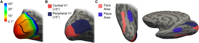
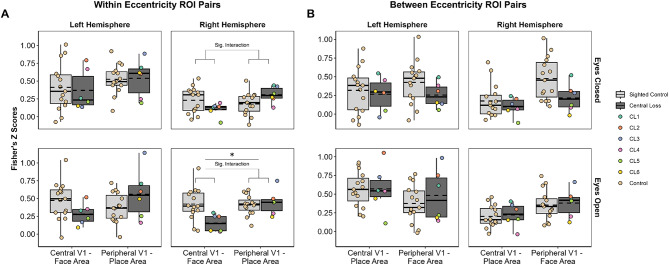
Individuals with central visual deficits exhibit atrophy of the visual cortex in regions representing the central visual field and show little or no functional response there. Information in the central and peripheral visual field appear to be represented preferentially in extrastriate regions that are selective to faces and places, respectively. We recruited individuals with bilateral macular degeneration (age-related or juvenile) and age-matched sighted controls. We used resting state fMRI (RS-fMRI) to examine functional connectivity between striate (V1) and extrastriate face and place selective areas as it allows better comparison between those with unaffected vision and those with visual loss, whose stimulus related signals are already known to differ from those of controls. Selective deficits emerged in our central loss group, showing reduced functional connectivity between regions with foveal biases (central V1-face area) compared to sighted controls, whereas no such difference emerged in the peripheral biased regions (peripheral V1-place area). This result was evident regardless of whether eyes were closed or open and fixating, but was only significant in the right hemisphere, supporting the functional lateralisation of face processing. This pilot study provides some evidence for reduced functional connectivity between foveal-biased visual areas in central vision loss, suggesting that communication within the posterior visual pathway may be selectively affected in partial vision loss. Functional connectivity differences did not appear to be driven by changes in viewing condition. RS-fMRI is a valuable tool that allows us to explore functional brain changes without the need for retinal input.
Keywords: Central visual deficits; Faces; Macular disease; Places; Visual cortex; fMRI.
© 2025. The Author(s).
Conflict of interest statement
Declarations. Ethics approval and consent to participate: This study followed the tenets of the Declaration of Helsinki with approval granted by the York Neuroimaging Centre (YNiC) Research, Ethics and Governance Committee. Consent for publication: Consent from all participants to include their anonymised data in publications was obtained. Competing interests: The authors declare no competing interests.
Figures
Similar articles
-
Reading aids for adults with low vision.Cochrane Database Syst Rev. 2018 Apr 17;4(4):CD003303. doi: 10.1002/14651858.CD003303.pub4. Cochrane Database Syst Rev. 2018. PMID: 29664159 Free PMC article.
-
Prophylactic non-steroidal anti-inflammatory drugs for the prevention of macular oedema after cataract surgery.Cochrane Database Syst Rev. 2016 Nov 1;11(11):CD006683. doi: 10.1002/14651858.CD006683.pub3. Cochrane Database Syst Rev. 2016. PMID: 27801522 Free PMC article.
-
Interhemispheric integration in the neural face perception network: Does stimulus location matter?Imaging Neurosci (Camb). 2025 May 29;3:IMAG.a.17. doi: 10.1162/IMAG.a.17. eCollection 2025. Imaging Neurosci (Camb). 2025. PMID: 40800977 Free PMC article.
-
Surgery for cataracts in people with age-related macular degeneration.Cochrane Database Syst Rev. 2017 Feb 16;2(2):CD006757. doi: 10.1002/14651858.CD006757.pub4. Cochrane Database Syst Rev. 2017. PMID: 28206671 Free PMC article.
-
Surgical interventions for bilateral congenital cataract in children aged two years and under.Cochrane Database Syst Rev. 2022 Sep 15;9(9):CD003171. doi: 10.1002/14651858.CD003171.pub3. Cochrane Database Syst Rev. 2022. PMID: 36107778 Free PMC article.
References
-
- Baseler Ha, Gouws A, Haak KV, Racey C, Crossland MD, Tufail A, Rubin GS, Cornelissen FW, Morland AB (2011) Large-scale remapping of visual cortex is absent in adult humans with macular degeneration. Nat Neurosci 14(5):649–655. 10.1038/nn.2793 - PubMed
MeSH terms
Grants and funding
LinkOut - more resources
Full Text Sources
Medical
