. 2025 Jul 3;21(7):e1013258.
doi: 10.1371/journal.ppat.1013258. eCollection 2025 Jul.

The non-structural protein of SFTSV activates NLRP1 and CARD8 inflammasome through disrupting the DPP9-mediated ternary complex

Affiliations

The non-structural protein of SFTSV activates NLRP1 and CARD8 inflammasome through disrupting the DPP9-mediated ternary complex

Pan-Pan Liu et al. PLoS Pathog. .

Abstract

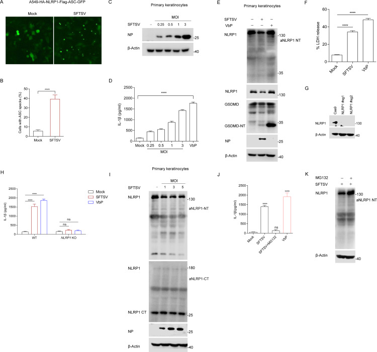
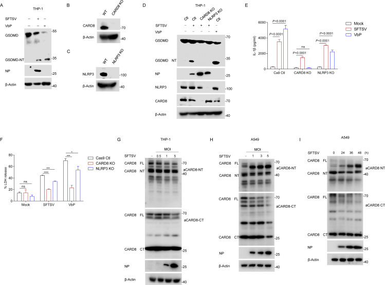
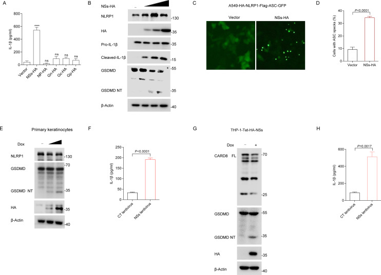
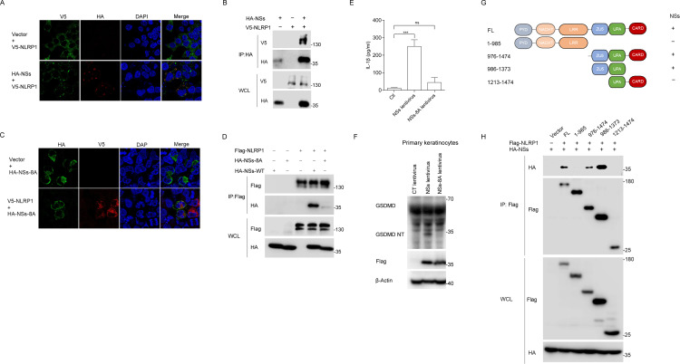
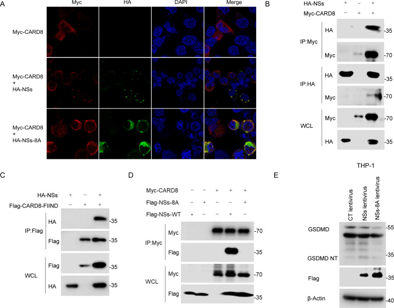
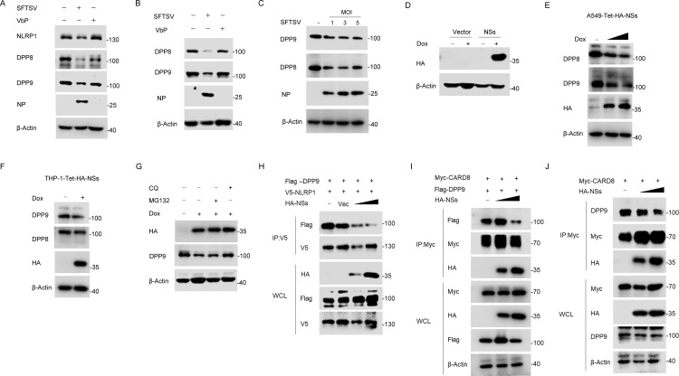
Inflammasomes function as immune-signaling platforms that were assembled following detection of pathogens. NLRP1 and CARD8 are related inflammasomes that use their C-terminal (CT) fragments containing a caspase recruitment domain (CARD) and the UPA domain to initiate the inflammasome. At rest, dipeptidyl peptidases 8 and 9 (DPP8/9) inhibit inflammatory CT by interacting with the function-to-find domain (FIIND) of NLRP1/CARD8 and forming an inhibitory NLRP1/CARD8-DPP9 ternary complex consisting of DPP9, full-length NLRP1/CARD8, and NLRP1/CARD8 CT. However, the specific triggers of NLRP1 and CARD8 have not yet been fully identified. Here, we report that a tick-borne bunyavirus SFTSV infection activates the NLRP1 inflammasome in primary keratinocytes and the CARD8 inflammasome in macrophages in a similar manner by targeting the ternary inhibitory complex, respectively. Mechanistically, SFTSV NSs interact with NLRP1 and CARD8 via their FIIND domains, suggesting that DPP8/9 are likely to compete for binding; on the other hand, NSs promote the degradation of DPP8 and DPP9. Both contribute to more efficient destabilization of the DPP8/9 ternary complex and release the activated CT. Moreover, CARD8 deletion promotes SFTSV replication. In conclusion, we found a novel mechanism of viral protein activation of NLRP1 and CARD8 by disrupting the DPP9-binding checkpoint.


Conflict of interest statement

The authors have declared that no competing interests exist.

Figures

Fig 1
Fig 1. SFTSV induces NLRP1 inflammasome activation in primary keratinocytes.
(A-B) ASC specks fluorescence microscopy images (A) and quantification (B) of A549-HA-NLRP1-Flag-ASC-GFP cells infected with SFTSV or without SFTSV (MOI = 0.5) for 24 h. Scale bar, 100 μm. (C-D) Detection of SFTSV NP (C) and IL-β production (D) in primary keratinocytes infected with SFTSV at different MOIs or stimulated with VbP (2 μM) for 24 h. (E-F) GSDMD cleavage (E) and LDH release (F) in primary keratinocytes infected with SFTSV (MOI = 1) or stimulated with VbP (5 μM) for 36 h. (G) Expression of NLRP1 in primary keratinocytes treated with lentivirus-mediated CRISPR-Cas9 and NLRP1-specific single-guide RNA (sgRNA) or the non-targeting sgRNA control. (H) IL-1β production in wild-type or NLRP1-deficient primary keratinocytes infected with SFTSV (MOI = 1) or stimulated with VbP (2 μM) for 24 h. (I) Primary keratinocytes were infected with SFTSV at different MOIs for 24 h, endogenous NLRP1 was detected using N-terminal and C-terminal antibodies with Western blot, respectively. (J-K) Primary keratinocytes were infected with SFTSV (MOI = 1) and treated with MG132 (2 μM) for 24 h, IL-1β release (J) in the cell supernatant was measured with ELISA; endogenous NLRP1 (K) was detected with Western blot. All data represent three independent experiments and presented as mean±s.d. *P < 0.05, **P < 0.01, ***P < 0.001, ****P < 0.0001, ns, not significant. For statistical analysis, one-way ANOVA in (D, J), two-tailed unpaired Student’s t-test in (B, F, H).
Fig 2
Fig 2. SFTSV activates the CARD8 inflammasome in macrophages.
(A) GSDMD cleavage in THP-1 cells infected with SFTSV (MOI = 1) or stimulated with VbP (2 μM) for 24 h. (B) Knockout of CARD8 in THP-1 cells identified with Western blot. (C) Knockout of NLRP3 in THP-1 cells identified with Western blot. (D-F) GSDMD cleavage (D), IL-1β production (E) and LDH release (F) in THP-1 WT, CARD8 KO and NLRP3 KO infected with SFTSV (MOI = 1) or stimulated with VbP (5 μM) for 24 h. (G) THP-1 cells were infected with SFTSV at different MOIs for 24 h, endogenous CARD8 was detected using N-terminal and C-terminal antibodies with Western blot, respectively. (H) A549 cells were infected with SFTSV at different MOIs for 24 h, endogenous CARD8 was detected using N-terminal and C-terminal antibodies with Western blot, respectively. (I) A549 cells were infected with SFTSV (MOI = 1) at indicated time, endogenous CARD8 was detected using N-terminal and C-terminal antibodies with Western blot, respectively. All data represent three independent experiments and presented as mean±s.d. *P < 0.05, **P < 0.01, ***P < 0.001, ****P < 0.0001, ns, not significant. For statistical analysis, two-tailed unpaired Student’s t-test in (E, F).
Fig 3
Fig 3. The role of SFTSV NSs in the activation of the NLRP1 and CARD8 inflammasome.
(A) ASC-caspase-1-pro-IL-1β HEK293T cells were transfected with indicated expression vector for 36 h. Supernatants were analyzed for IL-1β with ELISA. (B) ASC-caspase-1-pro-IL-1β HEK293T cells were transfected with plasmids NSs, production of p17 and GSDMD processing were detected with Western blot. (C-D) ASC specks fluorescence microscopy images (C) and quantification (D) of A549-HA-NLRP1-Flag-ASC-GFP cells transfected NSs-HA for 24 h. Scale bar, 100 μm. (E-F) GSDMD cleavage (E) and IL-1β (F) production in primary keratinocytes expressing Tet-HA-NSs and treated with doxycycline (1 μg/ml) for 48 h. (G-H) GSDMD cleavage (G) and IL-1β (H) production in THP-1 cells expressing Tet-HA-NSs and treated with doxycycline (1 μg/ml) for 48 h. All data represent three independent experiments and presented as mean±s.d. *P < 0.05, **P < 0.01, ***P < 0.001, ****P < 0.0001, ns, not significant. For statistical analysis, two-tailed unpaired Student’s t-test in (A, D, F, H).
Fig 4
Fig 4. SFTSV NSs interacts with NLRP1.
(A) Colocalization of NLRP1 and NSs in HEK293T cells co-transfected with V5-NLRP1 and HA-NSs for 48 h. Scale bar is 20 μm.(B) Co-IP assay between V5-NLRP1 and HA-NSs in HEK293T cells transfected with the indicated expression vectors for 48 h. (C) Colocation of NLRP1 and NSs-8A in HEK293T cells co-transfected with V5-NLRP1 and HA-NSs-8A for 48 h. Scale bar is 20 μm. (D) Co-IP assay between HA-NSs or HA-NSs-8A and Flag-NLRP1 in HEK293T cells transfected with the indicated expression vectors for 48 h. (E-F) IL-1β production (E) and GSDMD cleavage (F) in primary keratinocytes infected with lentiviruses expression Flag-NSs-WT or Flag-NSs-8A or carrying vector for 48 h. (G) Summary of mapping experiments to identify the NSs-binding domain in NLRP1. (H) Co-IP assay between NSs and NLRP1 domain truncation in HEK293T cells transfected with the indicated expression vectors for 48 h. All data represent three independent experiments and presented as mean±s.d. *P < 0.05, **P < 0.01, ***P < 0.001, ****P < 0.0001, ns, not significant. For statistical analysis, two-tailed unpaired Student’s t-test in (E).
Fig 5
Fig 5. SFTSV NSs interacts with CARD8.
(A) Colocalization of NSs, NSs-8A and NLRP1 in HEK293T cells co-transfected with HA-NSs, HA-NSs-8A and V5-NLRP1 for 48 h. Scale bar is 10 μm. (B) Co-IP assay between HA-NSs and Myc-CARD8 in HEK293T cells transfected with the indicated expression vectors for 48 h. (C) Co-IP assay between HA-NSs and Flag-CARD8-FIIND in HEK293T cells transfected with the indicated expression vectors for 48 h. (D) Co-IP assay between Flag-NSs or Flag-NSs-8A and Myc-CARD8 in HEK293T cells transfected with the indicated expression vectors for 48 h. (E) GSDMD cleavage in THP-1 cells infected with lentiviruses expression Flag-NSs-WT or Flag-NSs-8A or carrying vector for 48 h.
Fig 6
Fig 6. NSs impairs the interaction of NLRP1/CARD8 and DPP9.
(A) Primary keratinocytes were infected with SFTSV (MOI = 1) or stimulated with VbP (5 μM) for 36 h, endogenous DPP8 and DPP9 were detected with Western blot. (B) THP-1 cells were infected with SFTSV (MOI = 1) or stimulated with VbP (2 μM) for 24 h, endogenous DPP8 and DPP9 were detected with Western blot. (C) A549 cells were infected with different MOI SFTSV for 24 h, endogenous DPP8 and DPP9 were detected with Western blot. (D) A549 cells expressing Tet-HA-NSs were treated with doxycycline (1 μg/ml) for 48 h, and identified with Western blot. (E) A549-Tet-HA-NSs cells were treated with doxycycline (1 μg/ml) for 48 h, endogenous DPP8 and DPP9 were detected with Western blot. (F) THP-1-Tet-HA-NSs cells were treatment with doxycycline (1 μg/ml) for 48 h, endogenous DPP8 and DPP9 were detected with Western blot. (G) A549-Tet-HA-NSs cells were treatment with doxycycline (1 μg/ml) for 48 h and then treated with MG132 (10 μM) or CQ (50 μM) for 6 h before harvest. (H) Co-IP of NLRP1-DPP9 with NSs in HEK293T cells transfected with indicated expression vectors for 48 h. (I) Co-IP of CARD8-DPP9 with NSs in HEK293T cells transfected with indicated expression vectors for 48 h. (J) Co-IP of CARD8-DPP9 with NSs in HEK293T cells transfected with Myc-CARD8 and HA-NSs for 48 h.
Fig 7
Fig 7. CARD8 deletion promotes SFTSV replication.
(A-D) THP-1-Ctl, CARD8 KO, and NLRP3 KO were infected with SFTSV (MOI = 1) for 48 h, the mRNA levels (A) of SFSTV S, M, L segments were detected with RT-qPCR; (B) cells were stained with SFTSV NP and analyzed with immunofluorescence assays. Scale bar, 100 μm; (C) Expression of SFTSV NP was analyzed with Western blot; (D) Functional titers of SFTSV in Ctl, CARD8-KO and NLRP3-KO THP-1 cells measured by TCID50. (E-F) THP-1 cells were infected with SFTSV (MOI = 1) and treated with VbP (2 μM) for 24 h. (E) Expression of SFTSV NP was analyzed with Western blot; (F) cells were stained with SFTSV NP and analyzed with immunofluorescence assays. Scale bar, 100 μm. All data represent three independent experiments and presented as mean±s.d. *P < 0.05, **P < 0.01, ***P < 0.001, ****P < 0.0001, ns, not significant. For statistical analysis, two-tailed unpaired Student’s t-test in (A, D).

Similar articles

References

    1. Broz P, Dixit VM. Inflammasomes: mechanism of assembly, regulation and signalling. Nat Rev Immunol. 2016;16(7):407–20. doi: 10.1038/nri.2016.58 - DOI - PubMed
    1. Finger JN, Lich JD, Dare LC, Cook MN, Brown KK, Duraiswami C, et al. Autolytic proteolysis within the function to find domain (FIIND) is required for NLRP1 inflammasome activity. J Biol Chem. 2012;287(30):25030–7. doi: 10.1074/jbc.M112.378323 - DOI - PMC - PubMed
    1. Bauernfried S, Scherr MJ, Pichlmair A, Duderstadt KE, Hornung V. Human NLRP1 is a sensor for double-stranded RNA. Science. 2021;371(6528):eabd0811. doi: 10.1126/science.abd0811 - DOI - PubMed
    1. Taabazuing CY, Griswold AR, Bachovchin DA. The NLRP1 and CARD8 inflammasomes. Immunol Rev. 2020;297(1):13–25. doi: 10.1111/imr.12884 - DOI - PMC - PubMed
    1. D’Osualdo A, Weichenberger CX, Wagner RN, Godzik A, Wooley J, Reed JC. CARD8 and NLRP1 undergo autoproteolytic processing through a ZU5-like domain. PLoS One. 2011;6(11):e27396. doi: 10.1371/journal.pone.0027396 - DOI - PMC - PubMed

MeSH terms

LinkOut - more resources