. 2025 Jul 3;20(7):e0326897.
doi: 10.1371/journal.pone.0326897. eCollection 2025.

Semaglutide ameliorates diabetes-associated cognitive dysfunction in mouse model of type 2 diabetes

Affiliations

Semaglutide ameliorates diabetes-associated cognitive dysfunction in mouse model of type 2 diabetes

Yan Zhu et al. PLoS One. .

Abstract

Background: Type 2 diabetes mellitus (T2DM) is associated with cognitive dysfunction, which significantly impacts the quality of life. Semaglutide, a glucagon-like peptide-1 (GLP-1) receptor agonist, has shown potential neuroprotective effects. This study investigates the efficacy of semaglutide in ameliorating cognitive dysfunction in a mouse model of T2DM.

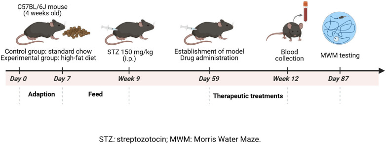
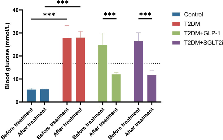
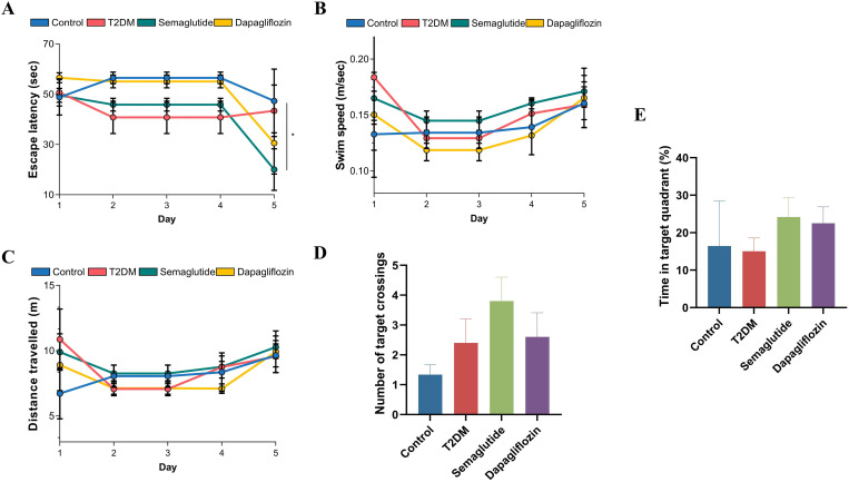
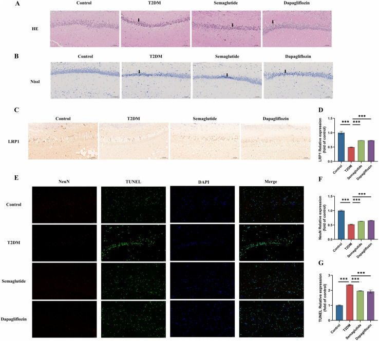
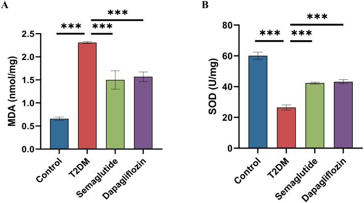
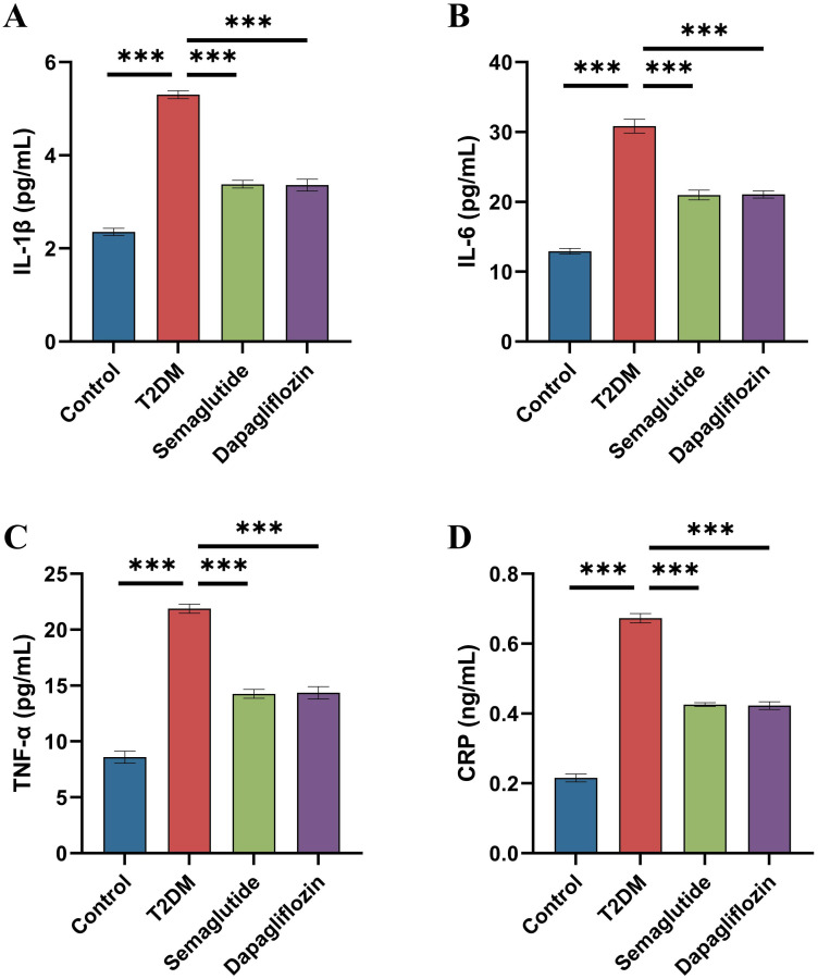
Methods: Male C57BL/6J mice were fed a high-fat diet for four weeks and received a single intraperitoneal injection of streptozotocin (150 mg/kg) to induce T2DM. All mice were divided into four groups: control, diabetes control (T2DM), semaglutide treatment (semaglutide, 0.1 mg/kg) and dapagliflozin treatment (dapagliflozin 1 mg/kg). Cognitive function was assessed using the Morris water maze (MWM) test. Histomorphological analysis of hippocampal tissues was performed using H&E and Nissl staining. Immunofluorescence was used to assess LRP1 expression and apoptosis. Biochemical analyses measured oxidative stress markers (SOD, MDA) and inflammatory cytokines (IL-1β, IL-6, TNF-α, CRP).

Results: Semaglutide treatment significantly reduced blood glucose levels in diabetic mice. In the MWM test, semaglutide-treated mice showed reduced escape latencies, indicating improved spatial learning and memory. Histomorphological analysis revealed preserved neuronal structure in the hippocampus with reduced neuronal damage and apoptosis in the semaglutide-treated group. Immunofluorescence showed increased LRP1 expression and decreased apoptosis. Biochemical analyses indicated that semaglutide reduced oxidative stress and inflammatory markers, further supporting its neuroprotective effects.

Conclusions: Semaglutide effectively ameliorates cognitive dysfunction in T2DM mice, likely through mechanisms involving the reduction of oxidative stress, inflammation, and neuronal apoptosis. These findings suggest that semaglutide has potential as a therapeutic agent for managing diabetes-associated cognitive decline. Further research, including long-term studies and clinical trials, is necessary to validate these findings and explore the broader applicability of semaglutide in treating cognitive impairments in diabetic patients.


Conflict of interest statement

The authors have declared that no competing interests exist.

Figures

Fig 1
Fig 1. Schematic diagram of the study design.
Fig 2
Fig 2. Effects of Semaglutide and Dapagliflozin on Blood Glucose Levels in T2DM Mice.
Blood glucose levels were measured before and after 4 weeks of treatment across four experimental groups: Control (blue), T2DM (red), T2DM+GLP-1 (green, semaglutide-treated), and T2DM+SGLT2i (purple, dapagliflozin-treated). Before treatment, all diabetic groups (T2DM, T2DM+GLP-1, and T2DM+SGLT2i) showed significantly elevated blood glucose levels compared to the Control group (***p < 0.001). After treatment, the Control group maintained normal glycemic levels, while the untreated T2DM group remained hyperglycemic. Both semaglutide and dapagliflozin treatments resulted in significant reductions in blood glucose levels compared to pre-treatment values and the untreated T2DM group (***p < 0.001). Data are presented as mean ± standard deviation (SD).
Fig 3
Fig 3. Assessment of cognitive function using the Morris water maze test.
(A) Escape latency (time to find the hidden platform) over five training days. The semaglutide group (green) showed progressively shorter escape latency compared to the T2DM group (red), with the most pronounced difference on day 5. The dapagliflozin group (yellow) showed improvement primarily on day 5. The control group (blue) maintained consistent performance throughout training. (B) Swim speed during the training days showed no significant differences between groups, indicating that motor function was not a confounding factor in cognitive assessment. (C) Distance traveled before finding the platform across training days, showing patterns similar to escape latency results, with the semaglutide group demonstrating improved performance compared to the untreated T2DM group. (D) Number of target crossings during the probe trial. The semaglutide group showed the highest number of platform location crossings, followed by the T2DM group, control group, and dapagliflozin group. (E) Time spent in the target quadrant during the probe trial. Both treatment groups (semaglutide and dapagliflozin) spent more time in the target quadrant compared to the control and T2DM groups, with the semaglutide group showing the highest percentage.Data are presented as mean ± standard deviation.Statistical significance is indicated as *p < 0.05.
Fig 4
Fig 4. Effects of Semaglutide and Dapagliflozin on Neuronal Integrity, Apoptosis, and LRP1 Expression in the Hippocampal CA1 Region of T2DM Mice.
(A) Hematoxylin and eosin (H&E) staining of hippocampal CA1 region shows preserved neuronal structure in the control, semaglutide and dapagliflozin groups compared to the T2DM group. Black arrows indicate nuclear pyknosis (condensed, shrunken nuclei) in the T2DM group, suggesting neuronal damage. (B) Nissl staining further confirms the preservation of neuronal integrity in control and treated groups, with the T2DM group showing reduced Nissl substance. Black arrows indicate areas of neuronal loss and structural disruption in the hippocampal CA1 region. (C) Immunohistochemical staining for LRP1 (Low-density lipoprotein receptor-related protein 1) reveals reduced expression in the T2DM group, with restoration observed in both semaglutide and dapagliflozin-treated groups. (D) Quantitative analysis of LRP1 expression shows significantly decreased levels in the T2DM group compared to control (***p < 0.001), while both semaglutide and dapagliflozin treatment significantly restored LRP1 expression compared to the T2DM group (***p < 0.001). (E) Double immunofluorescence staining for NeuN (neuronal marker, red) and TUNEL (apoptosis marker, green), with DAPI nuclear counterstain (blue). The merged images show substantially increased neuronal apoptosis in the T2DM group compared to control, with both semaglutide and dapagliflozin treatments reducing apoptotic signals. (F) Quantitative analysis of NeuN-positive cells demonstrates significant neuronal loss in the T2DM group compared to control (***p < 0.001), with both treatments preserving neuronal populations compared to the untreated T2DM group (***p < 0.001). (G) Quantitative analysis of TUNEL-positive cells confirms significantly increased apoptosis in the T2DM group compared to control (***p < 0.001), with both semaglutide and dapagliflozin treatments significantly reducing neuronal apoptosis (***p < 0.001). Data are presented as mean ± standard deviation (SD).
Fig 5
Fig 5. Effects of Semaglutide and Dapagliflozin on Oxidative Stress Markers in the Hippocampus of T2DM Mice.
(A) MDA levels were significantly higher in the T2DM group compared to the control group (***p < 0.001). Both semaglutide and dapagliflozin treatments significantly reduced MDA levels compared to the T2DM group (***p < 0.001), with no significant difference between the treated groups. (B) SOD activity was significantly lower in the T2DM group compared to the control group (***p < 0.001). Treatment with semaglutide and dapagliflozin significantly increased SOD activity compared to the T2DM group (***p < 0.001), with no significant difference between the treated groups. Data are presented as mean ± standard deviation (SD). Statistical significance is indicated as ***p < 0.001.
Fig 6
Fig 6. Effects of Semaglutide and Dapagliflozin on Inflammatory Markers in T2DM Mice.
(A) IL-1β levels were significantly elevated in the T2DM group compared to the control group (***p < 0.001). Both semaglutide and dapagliflozin treatments significantly reduced IL-1β levels compared to the T2DM group (***p < 0.001), with no significant difference between the treated groups. (B) IL-6 levels followed a similar pattern, with significant reductions in the semaglutide and dapagliflozin groups compared to the T2DM group (***p < 0.001), and no significant difference between the two treatments. (C) TNF-α levels were also significantly higher in the T2DM group compared to the control group (***p < 0.001), with both treatments reducing TNF-α levels significantly (***p < 0.001), and no significant difference between the semaglutide and dapagliflozin groups. (D) CRP levels were elevated in the T2DM group, with significant reductions observed in the semaglutide and dapagliflozin groups compared to the T2DM group (***p < 0.001), and no significant difference between the treated groups. Data are presented as mean ± standard deviation (SD). Statistical significance is indicated as ***p < 0.001.

Similar articles

References

    1. GBD 2021 Diabetes Collaborators. Global, regional, and national burden of diabetes from 1990 to 2021, with projections of prevalence to 2050: a systematic analysis for the Global Burden of Disease Study 2021. Lancet. 2023;402(10397):203–34. doi: 10.1016/S0140-6736(23)01301-6 - DOI - PMC - PubMed
    1. Ali MK, Pearson-Stuttard J, Selvin E, Gregg EW. Interpreting global trends in type 2 diabetes complications and mortality. Diabetologia. 2022;65(1):3–13. doi: 10.1007/s00125-021-05585-2 - DOI - PMC - PubMed
    1. Biessels GJ, Staekenborg S, Brunner E, Brayne C, Scheltens P. Risk of dementia in diabetes mellitus: a systematic review. Lancet Neurol. 2006;5(1):64–74. doi: 10.1016/S1474-4422(05)70284-2 - DOI - PubMed
    1. Biessels GJ, Despa F. Cognitive decline and dementia in diabetes mellitus: mechanisms and clinical implications. Nat Rev Endocrinol. 2018;14(10):591–604. doi: 10.1038/s41574-018-0048-7 - DOI - PMC - PubMed
    1. Su J, Sun G, An J, Ao Y, Li J, Shen Z, et al. Efficacy and safety of the integration of traditional Chinese medicine and western medicine in the treatment of diabetes-associated cognitive decline: a systematic review and meta-analysis. Front Pharmacol. 2023;14:1280736. doi: 10.3389/fphar.2023.1280736 - DOI - PMC - PubMed

MeSH terms