. 2025 Jul 3:blood.2025028935.
doi: 10.1182/blood.2025028935. Online ahead of print.

Restoring p53 wild-type conformation in TP53-Y220C-mutant acute myeloid leukemia

Affiliations

Restoring p53 wild-type conformation in TP53-Y220C-mutant acute myeloid leukemia

Bing Z Carter et al. Blood. .

Abstract

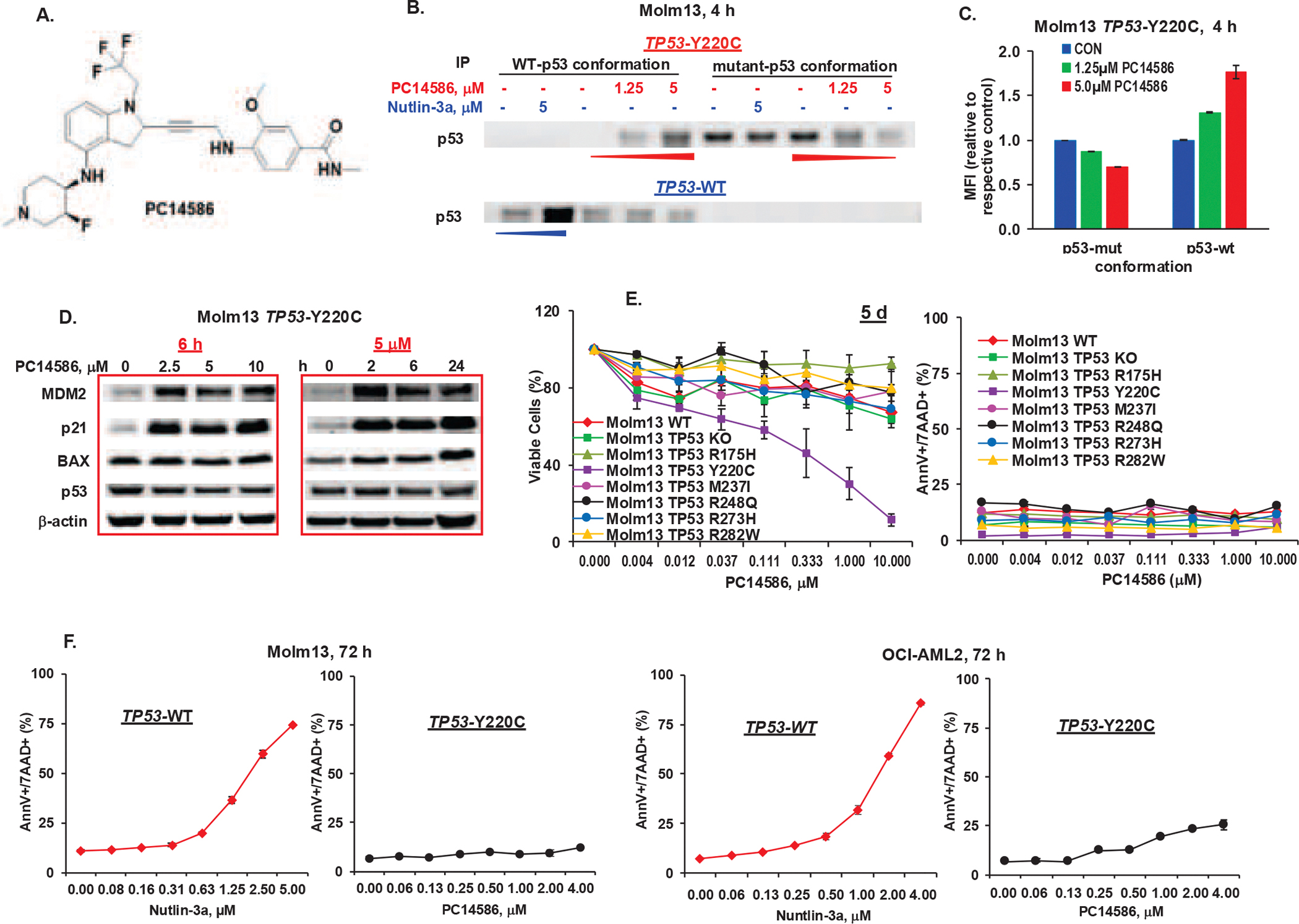
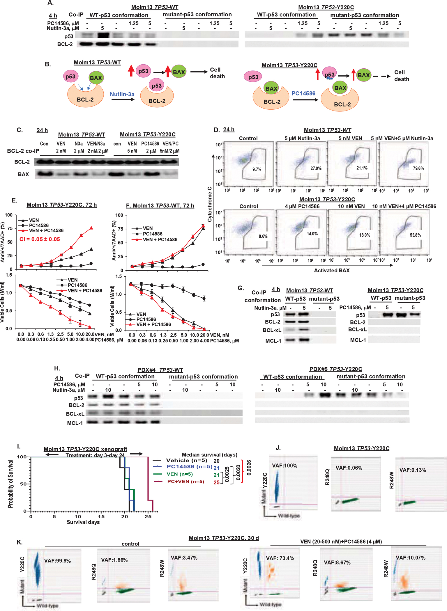
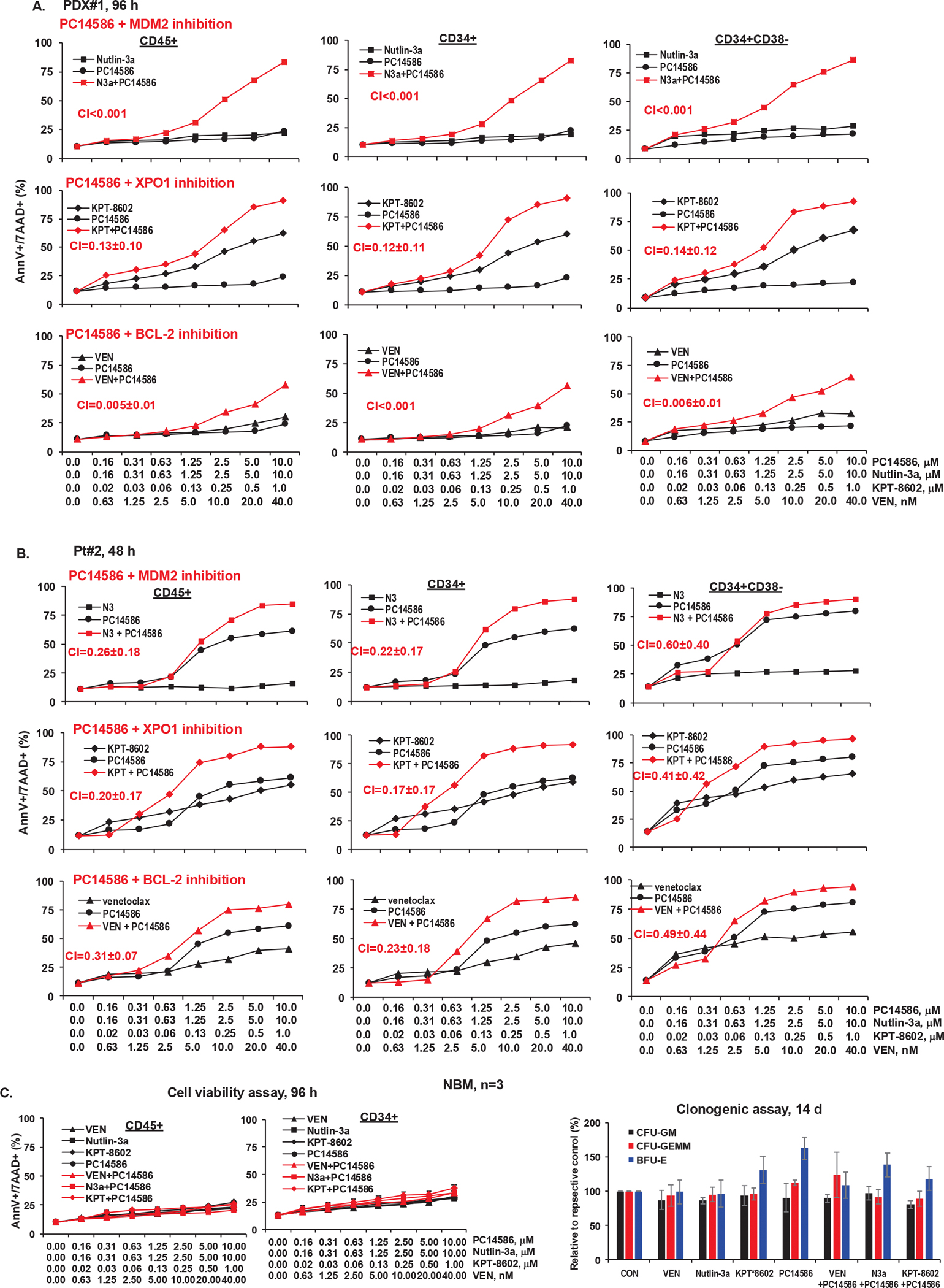
TP53-Y220C is a recurrent hotspot mutation in cancers and leukemias. It is observed predominantly in acute myeloid leukemia (AML)/myelodysplastic syndromes among hematological malignancies and is associated with poor outcome. The mutation creates a structural pocket in the p53 protein. PC14586 (rezatapopt) is a small molecule designed to bind to this pocket and thus restore a p53-wild type (p53-WT) conformation. We demonstrate that PC14586 converts p53-Y220C into a p53-WT conformation and activates p53 transcriptional targets, but surprisingly induces limited/no apoptosis in TP53-Y220C AML. Mechanistically, MDM2 induced by PC14586-activated conformational p53-WT and the nuclear exporter XPO1 reduce the transcriptional activities of p53, which are fully restored by inhibition of MDM2 and/or XPO1. Importantly, p53-WT protein can bind to BCL-2, competing with BAX in the BH3 binding pocket of BCL-2 and also binds to BCL-xL and MCL-1. However, such binding by PC14586-activated conformational p53-WT is not detected. Pharmacological inhibition of the BCL-2/BAX interaction with venetoclax fully compensates for this deficiency, induces massive cell death in AML cells and stem/progenitor cells in vitro and prolongs survival of TP53-Y220C AML xenografts in vivo. Collectively, we identified transcription-dependent and -independent mechanisms that limit the apoptogenic activities of reactivated conformational p53-WT and suggest approaches to optimize apoptosis induction in TP53-mutant leukemia. A clinical trial of PC14586 in TP53-Y220C AML/myelodysplastic syndromes has recently been initiated (NCT06616636).


Conflict of interest statement

Conflict of interest disclosure: B.Z.C. and M.A. received research funding from PMV, A.P.K. and M.V.P. are employees of PMV, and A.L. is on the board directors of PMV and a consultant for the science of p53 and for Chugai Pharma for monoclonal antibodies.

Figures

Figure 1.
Figure 1.. PC14586 stabilizes the p53-WT conformation, induces the activation of p53 signaling, and has antileukemia activity in TP53-Y220C AML cells.
A. Structure of PC14586. B. TP53-Y220 and TP53-WT Molm13 cells were treated with the indicated concentrations of PC14586 or nutlin-3a for 4 h. The p53-WT and p53-mutant conformations were determined by IP with conformation-selective antibodies followed by Western blotting. C. TP53-Y220 Molm13 cells were treated with indicated concentrations of PC14586 for 4 h. The p53-WT and p53-mutant conformations were determined by flow cytometry with conformation-selective antibodies. D. TP53-Y220 Molm13 cells were treated with the indicated concentrations of PC14586 for 6 h or with 5 μM PC14586 for the indicated times. Protein levels were determined by Western blotting. E. Isogenic Molm13 cells with TP53-WT, KO, various mutations were treated with the indicated concentrations of PC14586 for 5 d. Viable cells and cell death were determined by flow cytometry. F. TP53-WT and TP53-Y220C Molm13 or OCI-AML2 cells were treated with the indicated concentrations of nutlin-3a or PC14586, respectively, for 72 h. Cell death was determined by flow cytometry. Mut, mutant; d, day; h, hour; MFI, mean fluorescence intensity; AnnV, annexin V; 7AAD, 7-aminoactinomycin D. Cell death and viability experiments were performed in triplicate; results were expressed as means±standard errors of the means.
Figure 2.
Figure 2.. PC14586 combined with MDM2 or XPO1 inhibition further upregulates p53 transcriptional activities and target proteins, synergistically induces cell death, and suppresses cell growth in TP53-Y220C cells.
TP53-WT or TP53-Y220C Molm13 cells were treated with PC14586 (4 μM), nutlin-3a (5 μM), or both for 4 h and subjected to RNA-seq (n=3) (A-C) and Western blotting (D). A. PCA of the treated cells. B. Heatmap of the DGE analysis (adjusted P ≤0.05, log2FC≥ 1, and baseMean ≥1000). C. Integrated Hallmark GSEA pathways (Adj P ≤0.001) with the x-axis representing different comparisons, y-axis representing various pathways, dot color representing −log10 P adj) x direction (i.e., red color indicates upregulation direction, while blue downregulation direction), and dot size representing absolute value of normalized enrichment scores. D. Validation of p53 target proteins by Western blotting. E. TP53-Y220C Molm13 cells were treated with PC14586, nutlin-3a or both for 72 h. Cell death and viable cells were determined by flow cytometry. F. TP53-Y220C Molm13 cells were treated with PC14586 (4 μM), KPT-8602 (200 nM), or both for 24 h. p53 localization and its target proteins were determined by Western blotting. G. TP53-Y220C Molm13 cells were treated with PC14586, KPT-8602, or both for 72 h. H-I. TP53-Y220C (H) or -WT (I) Molm13 cells were treated with PC14586, nutlin-3a, KPT-8602, or various combinations for 72 h. Cell death and viable cells were determined by flow cytometry. DEGs, differentially expressed genes; con, control; PC, PC14586; N3a, nutlin-3a; MWM, molecular weight marker; M/ml, million/ml; AnnV, annexin V; 7AAD, 7-aminoactinomycin D. Cell death and viability experiments were performed in triplicate; results were expressed as means±standard errors of the means.
Figure 3.
Figure 3.. PC14586-reacativated p53 does not bind to BCL-2 proteins and PC14586 synergizes with VEN.
A. TP53-WT or -Y220C Molm13 cells were treated with nutlin-3a (5 μM) or PC14586 (4 μM) for 4 h. p53-BCL-2 interaction was determined by pulling down p53 with antibodies selective for the p53-WT- or -mutant conformation in the treated cells, followed by Western blotting for p53 or BCL-2. B. The proposed mechanism of action of PC14586 in TP53-Y220C cells. Nutlin-3a in TP53-WT cells is used as a control. C and D. TP53-WT Molm13 cells were treated with VEN, nutlin-3a or both and TP53-Y220C cells were treated with VEN, PC14586, or both at the indicated doses for 24 h. BCL-2 was pulled down by IP and BCL-2 bound BAX (co-IP) were determined by Western blotting (C). BAX activation and cytochrome C levels were determined by flow cytometry (D). E-F. TP53-Y220C (E) or -WT (F) Molm13 cells were treated with PC14586, VEN, or both for 72 h. Cell death and viable cells were determined by flow cytometry. G-H. TP53-WT or -Y220C Molm13 cells (G) or PDX cells (H) were treated with nutlin-3a or PC14586 for 4 h. p53-BCL-2, -BCL-xL, or -MCL-1 interaction was determined by co-IP followed by Western blotting. I. Survival curves for NSG mice bearing TP53-Y220C Molm13 xenografts and treated with PC14586, VEN, or both. J. Detection of TP53 mutations by ddPCR in Molm13 TP53-Y220C cells. K. Detection of TP53 mutations by ddPCR in TP53-Y220C Molm13 cells treated in vitro with PC14586 (4 μM) plus VEN (initially 20 nM and increased to 500 nM over time) or in untreated control cells for 30 days. Con, control; PC, PC14586; M/ml, million/ml; d, day; AnnV, annexin V; 7AAD, 7-aminoactinomycin D. Cell death and viability experiments were performed in triplicate; results were expressed as means±standard errors of the means.
Figure 4.
Figure 4.. PC14586 with concomitant MDM2, XPO1, or BCL-2 inhibition synergistically induces cell death in AML blasts and stem/progenitor cells from patients with the TP53-Y220C mutation but has limited toxicity in NBM cells and stem/progenitor cells.
A-C, Cells from PDX#1 (A), Pt#2 (B), and NBM (n=3) (C) were treated with PC14586 alone or in combination with the indicated concentrations of nutlin-3a, KPT-8602, or VEN for the indicated times. Cell death was determined by flow cytometry in CD45+, CD34+, and CD34+CD38 cells. For NBM cells, cell death (left) and colony forming units (CFU) of cells treated with VEN (40 nM), nutlin-3a (10 μM), KPT-8602 (1 μM), PC14586 (10 μM), and each agent in combinations with PC14586 (right) are shown. N3a or N3, nutlin-3a; KPT, KPT-8602; AnnV, annexin V; 7AAD, 7-aminoactinomycin D.
Figure 5.
Figure 5.. PC14586 in combination with RG7388 has therapeutic efficacy in a PDX model in vivo.
NSG mice harboring PDX cells with TP53-Y220C and TP53-P151A mutations and other co-mutations were treated with vehicle, PC14586, RG7388, or both from d-38 to d-129 (RG7388 was stopped from d-97 to d-111). A. Circulating blasts after 4-week (left), 6-week (middle), or 8-weerk (right) treatment were determined by flow cytometric measurement of huCD45+ cells in PB. B. Spleen sizes (left) and weights(right) after 4-week treatment. C. BM blasts after 4-week treatment. D. Mouse survival. E-G. CyTOF analysis of BM cells collected after 4-week treatment. E. Treatment effects on various cell populations. F. Cell clustering based on cell surface markers (left and middle) and the effects of treatment on selected cell populations (right). G. tsne blots illustrate the effects of treatment on huCD45+ cells and p53-mutant in huCD45+ cells. RG, RG7388; PC, PC14586; d, day; Pop, population.
Figure 6.
Figure 6.. ScDNA-seq of BM cells collected from vehicle- and PC14586-treated mice at moribund stage.
NSG mice harboring PDX cells with TP53-Y220C and TP53-P151A mutations and other co-mutations (#1) were treated with vehicle or PC14586. BM cells were collected from moribund mice in each group and subjected to scDNA-seq using the MissionBio platform. A. Heatmap showing the frequencies of TP53-Y220C, TP53-P151A, NRAS-Q61R, and NRAS-Q61L mutations in BM cells from vehicle- or PC14586-treated mice. Arrows indicate whether the mutations in PC14586-treated mice were increased or decreased compared to vehicle-treated mice. B. Characterizations of clones. C. Fish plot showing clonal evolution (left) and clonal distribution (right).

Similar articles

References

    1. Hou HA, Chou WC, Kuo YY, et al. TP53 mutations in de novo acute myeloid leukemia patients: longitudinal follow-ups show the mutation is stable during disease evolution. Blood Cancer J. 2015;5:e331. - PMC - PubMed
    1. Stengel A, Kern W, Haferlach T, Meggendorfer M, Fasan A, Haferlach C. The impact of TP53 mutations and TP53 deletions on survival varies between AML, ALL, MDS and CLL: an analysis of 3307 cases. Leukemia. 2017;31(3):705–711. - PubMed
    1. Kadia TM, Jain P, Ravandi F, et al. TP53 mutations in newly diagnosed acute myeloid leukemia: Clinicomolecular characteristics, response to therapy, and outcomes. Cancer. 2016;122(22):3484–3491. - PMC - PubMed
    1. Short NJ, Montalban-Bravo G, Hwang H, et al. Prognostic and therapeutic impacts of mutant TP53 variant allelic frequency in newly diagnosed acute myeloid leukemia. Blood Adv. 2020;4(22):5681–5689. - PMC - PubMed
    1. DiNardo CD, Jonas BA, Pullarkat V, et al. Azacitidine and Venetoclax in Previously Untreated Acute Myeloid Leukemia. N Engl J Med. 2020;383(7):617–629. - PubMed

Associated data