. 2025 Dec;14(1):2529889.
doi: 10.1080/22221751.2025.2529889. Epub 2025 Jul 28.

Chlorinated biscoumarins inhibit chikungunya virus replication in cell-based and animal models

Affiliations

Chlorinated biscoumarins inhibit chikungunya virus replication in cell-based and animal models

Chidinma Nelson Orji et al. Emerg Microbes Infect. 2025 Dec.

Abstract

Biscoumarin derivatives were evaluated for antiviral activity against chikungunya virus (CHIKV), a re-emerging mosquito-borne alphavirus with no approved treatment. Compounds 3 and 4 demonstrated potent in vitro antiviral efficacy, with EC₅₀ values of 2.85 ± 0.42 µM and 3.08 ± 0.45 µM (SI > 20) for compound 3 in Vero and HEK293 cells, respectively. Compound 4 showed comparable potency in Vero cells but was less effective in HEK293 cells. Time-of-addition and replicon assays suggested that both compounds act at a post-entry step, likely inhibiting viral RNA replication. In vivo, a single oral dose of 250 mg/kg was well tolerated in mice and rats, with no signs of acute hepatorenal toxicity and favourable pharmacokinetic profiles. Compound 3 & 4 significantly reduced tissue viral loads within 24 hours; however, their antiviral effect diminished after the drug was cleared from circulation. Due to concerns about potential cumulative toxicity, repeated administration was avoided. Preliminary mechanistic studies indicated moderate inhibition of the viral nsP1 methyltransferase and suggested possible involvement of host pathways. These findings highlight biscoumarin derivatives - particularly compound 3 - as promising antiviral candidates against CHIKV, meriting further optimization and investigation into their mechanisms of action.

Keywords: Biscoumarin; chikungunya virus; efficacy; methyltransferase; mice; molecular docking.


Conflict of interest statement

No potential conflict of interest was reported by the author(s).

Figures
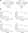
Figure 1.
Figure 1.
Antiviral activity of chlorinated biscoumarins against CHIKV in mammalian cells. (a) Chemical structures of compounds 3 and 4. (b) Dose – response curves showing CHIKV inhibition in Vero cells treated with compounds 3 and 4. Viral titres were measured by plaque assay at 24 hpi, and EC₅₀ values were calculated using non-linear regression. Selectivity indices (SI) were determined as the ratio of CC₅₀ to EC₅₀. (c) Dose-response curves showing CHIKV inhibition in HEK-293 cells treated with compounds 3 and 4 under identical conditions. Data are presented as mean ± SEM from three independent experiments performed in triplicate.
Figure 2.
Figure 2.
Compounds 3 and 4 inhibit CHIKV replication at a post-entry stage. (a) Schematic overview of the attachment inhibition assay. Vero cells were treated with compounds 3 or 4 (10 µM) during different phases of infection: pre-treatment (cells treated before virus exposure), pre-incubation (virus treated before cell exposure), co-incubation (virus and cells exposed to compound simultaneously), post-incubation (compound added after viral entry), and throughout (compound present continuously). (b) CHIKV inhibition (%) for each treatment condition was determined by plaque assay from culture supernatants at 24 h post-infection (hpi). Bars represent mean ± SD from triplicate wells. (c) Schematic of Time-of-Addition (TOA) and Time-of-Removal (TOR) assays. In TOA, compounds were added at various time points post-infection and maintained until 24 hpi. In TOR, compounds were added at 0 hpi and removed at designated time points. (d, e) CHIKV titres measured by plaque assay following TOA and TOR assays for compounds 3 (e) and 4 (e), showing a critical window of drug activity between 0–9 hpi. (f) Schematic of the CHIKV replicon construct encoding luciferase (Luc) and ZsGreen reporters, used to assess viral RNA replication. (g, h) Dose-response curves of compounds 3 (g) and 4 (h) in BHK-21 cells stably expressing the CHIKV replicon. Luciferase activity was measured at 24 h post-treatment to calculate EC₅₀ values. Data are shown as mean ± SD from three independent experiments.
Figure 3.
Figure 3.
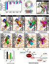
CHIKV methyltransferase binding and activities (a) in silico pan-docking of compounds 3 and 4 to viral proteins compared to their respective native inhibitors (green dash lines) (b, c) Binding poses, energy, and interacting residues of substrate (SAM), native inhibitor (Sinefungin), compounds 3 and 4 (d) in vitro methyltransferase assay (e) schematic diagram of alphavirus capping mechanism.
Figure 4.
Figure 4.
Assessment of acute oral toxicity of compounds 3 and 4 in mice. (a) Schematic representation of the single-dose toxicity study. C57BL/6 mice (5–6 per group) received a single oral dose of compounds 3 or 4 at 250 or 500 mg/kg. Blood samples were collected on days 1, 3, and 7 for liver (ALT) and kidney (creatinine) function tests. (b) Daily activity scores of mice monitored over a 7-day period following single-dose administration. (c, d) Serum levels of alanine transaminase (ALT, c) and creatinine (d) were measured on days 1, 3, and 7. (e) Schematic of the multiple-dose toxicity study. Compound 3 was administered orally at 250 mg/kg twice daily (BID) for 3 days. Ribavirin (50 mg/kg BID) and vehicle were used as controls. (f) Activity scores of mice during the multiple-dose study. (g, h) ALT (g) and creatinine (h) levels were measured on day 3 after the final dose.
Figure 5.
Figure 5.
Plasma-concentration time profile of compounds 3 and 4 in mice and rats (a, b) A scheme of experiment in mice (a) and rats (b). Time points used for collection of plasma samples are indicated by arrows pointing up. (c, d) Results of plasma-concentration time profile of 3 and 4 were plotted in means and standard deviation (n = 3), comparing between mice and rats. The EC50 and EC90 levels of 3 and 4 are marked with dotted lines. (e) The ALT and Cr levels were evaluated in rats at 24, 48, and 72 h after compounds’ administration.
Figure 6.
Figure 6.
In vivo efficacy assessment of compounds 3 and 4 in a CHIKV-induced arthritis mouse model. (a) Schematic of the experimental design. C57BL/6 mice (n = 18/group) were infected with CHIKV (5 × 107 PFU) via footpad injection and treated once daily with compound 3, compound 4 (250 mg/kg), ribavirin (50 mg/kg B.I.D), vehicle, or left untreated. An uninfected group served as a negative control (n = 6). Sampling of 6 mice/group was performed on days 1–4 post-infection. (b, c) Changes in foot length and width relative to day 0 (%). (d) Changes in body weight relative to day 0 (%). (e) Activity scores measured daily post-infection. (f) CHIKV titres in footpad tissues determined by plaque assay. (g) CHIKV titres in plasma determined by plaque assay. (h, i) CHIKV RNA levels in footpad tissues (h) and plasma (i) determined by RT-qPCR. Dashed line represents limit of detection (LOD). Each dot represents an individual mouse; horizontal lines indicate group means and standard deviation. Statistical significance was assessed using multiple paired t-test, Mann-Whitney test *p < 0.05.
Figure 7.
Figure 7.
H & E pathology of footpad samples on day 1, 2, and 3 after administering a single dose of 250 mg/kg compounds 3 or 4. Ribavirin was used as a positive control (50 mg/kg, BID, IP, 6 doses). The line represents a 100 µm scale. Representative H&E-stained sections of footpad tissue collected on days 1–3 post-infection. Treatment groups include compound 3 (250 mg/kg), compound 4 (250 mg/kg), ribavirin (50 mg/kg), and vehicle. Additional controls include CHIKV-infected mice before treatment (day 1), CHIKV-infected untreated mice (day 3), and uninfected mice (day 3). Tissue sections were examined for inflammatory cell infiltration and structural changes.

References

    1. Bartholomeeusen, K, et al. Chikungunya fever. Nat Rev Dis Primers. 2023;9(1):17. doi: 10.1038/s41572-023-00429-2 - DOI - PMC - PubMed
    1. Mourad, O, Makhani L, Chen LH.. Chikungunya: an emerging public health concern. Curr Infect Dis Rep. 2022;24(12):217–228. doi: 10.1007/s11908-022-00789-y - DOI - PMC - PubMed
    1. Chantasrisawad, N, et al. Characterization of clinical and biologic manifestations of chikungunya among children in an urban area, Thailand: a retrospective cohort study. Pediatr Infect Dis J. 2025;44:p e60–e62. doi: 10.1097/INF.0000000000004542 - DOI - PubMed
    1. Roongaraya, P, Boonyasuppayakorn S.. Chikungunya vaccines: an update in 2023. Asian Pac J Allergy Immunol. 2023;41(1):1–11. - PubMed
    1. Mullard, A. FDA approves first chikungunya vaccine. Nat Rev Drug Discov. 2024;23:8. doi: 10.1038/d41573-023-00201-x - DOI - PubMed

MeSH terms