. 2025 Jul 4;16(1):6147.
doi: 10.1038/s41467-025-60986-w.

The mechanistic basis for interprotomer deglycosylation of antibodies by corynebacterial IgG-specific endoglycosidases

Affiliations

The mechanistic basis for interprotomer deglycosylation of antibodies by corynebacterial IgG-specific endoglycosidases

Diego E Sastre et al. Nat Commun. .

Abstract

Corynebacterium diphtheriae clade species secrete single-domain endo-β-N-acetylglucosaminidases (ENGases) that specifically bind to human IgG antibodies and hydrolyze their N297-linked glycans. Here, we define the molecular mechanisms of IgG-specific deglycosylation for the entire family of corynebacterial IgG-specific ENGases, including but not limited to CU43 and CM49. By solving the crystal structure of CU43 in a 1:1 complex with the IgG1 Fc region, combined with targeted and saturation mutagenesis analysis and activity measurements using engineered antibodies, we establish an inter-protomeric mechanism of recognition and deglycosylation of IgG antibodies. Using in silico modeling, small-angle X-ray scattering and saturation mutagenesis we determine that CM49 uses a unique binding site on the Fc region, to process N297-linked glycans. Moreover, we demonstrate that CU43 treatment is highly effective in abrogating Fc effector functions in humanized mouse models, while preserving the neutralizing capacity of anti-influenza IgG antibodies, thereby conferring protection against lethal influenza challenge.


Conflict of interest statement

Competing interests: D.E.S., S.B, J.D., J.V.R. and E.J.S. are inventors on a provisional patent application filed with the United States Patent and Trademark Office by Emory University relevant to the work in this manuscript. All other authors declare they have no competing interests.

Figures

Fig. 1
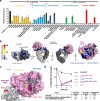
Fig. 1. Corynebacterial IgG-specific CU43 forms a complex with Fc with 1:1 stoichiometry.
a Schematic representation of domain architecture of IgG-specific ENGases (EndoS, EndoS2) and corynebacterial ENGase CU43. SP=signal peptide. b SEC-MALS analysis of CU43i-Fc complex in 2:1 molar ratio (upper panel) and the indicated fractions from SEC were loaded into SDS-PAGE in non-reducing (NR) conditions. Assays were performed in two independent replicates, and consistent results were observed. Blue squares represent GlcNAc, red triangles represent Fucose, Green circles represent Mannose and yellow circles represent Galactose. c AUC analysis of CU43i, Fc CT and CU43-Fc 1:1 complex. Samples in PBS were centrifuged at 45,400 xg. Curves represent fits to the continuous c(s) distribution model in SEDFIT. Res.=residuals. d SAXS scattering curve of CU43i, Fc-IgG1 and CU43i-Fc. eP(r) functions distributions of CU43i, Fc-IgG1 and CU43i-Fc complex. f Normalized Kratky plot CU43i, Fc-IgG1 and CU43i-Fc complex. g Ab initio modeling with GASBOR of the envelope of CU43i and superimposition of CU43i (PDB code: 8URA) crystal structure. h Ab initio modeling with GASBOR of the envelope of IgG1(Fc) and superimposition of Fc-IgG1 X-ray crystal structure (PDB code: 1H3X). i Ab initio modeling with GASBOR of the envelope of CU43i-Fc and superimposition of AF3 CU43i (orange)-Fc (gray) complex predicted model. Schematic figures in panels 1a and 1b were created in BioRender. Sastre, D. (https://BioRender.com/8gujq6o) and Sastre, D. (https://BioRender.com/uh09kf7) respectively. Source data are provided as a Source Data file.
Fig. 2
Fig. 2. Structural basis of human IgG-specific deglycosylation by CU43.
a Two views of cartoon representation of the crystal structure of CU43i-FcE382S crystal structure at 3.6 Å resolution (PDB code: 8URO). G0F N-glycan was docked into CU43i. b Surface representation of CU43i-FcE382S with glycan docked showing the distance to N297 from each Fc protomer. c Deep view of protein-protein interface residues of CU43i and FcE382S complex represented as sticks. d Two views of crystal structure of CU43i-FcE382S complex representing B-factor values. e Surface electrostatic potential of CU43 and Fc IgG1 obtained using (https://server.poissonboltzmann.org/pdb2pq). N-glycan G0F was docked in CU43i structure.
Fig. 3
Fig. 3. Targeted mutagenesis corroborates the IgG-specific deglycosylation mechanism of CU43.
a ENGase activity of CU43 WT and mutant variants against Fc WT or Fc mutant variants. Incubation of CU43 WT against Fc WT was considered 100 % of activity (1:500 enzyme:substrate molar ratio). Data is presented as mean values from independent samples ± SD (n = 3). Mean values were obtained by globally fitting the different kinetic traces data using Kintek Global Kinetic Explorer. b Surface representation of CU43i-Fc structure color-coded according to the relative activity of each mutant variant from panel A. Protomer B on CU43-FcE382S structure was replaced for a copy of Fc from PDB (code: 5JIH) to show all mutated residues. N-glycan G0F was docked into CU43i structure. c Surface representation of CU43 and Fc (protomer A) with a G0F glycan docked, in which are colored the key residues involved in the protein-protein interface of CU43 with Fc and the residues involved in the glycan binding site that were used in the in vivo assays indicated in (d). GlcNAc is colored in blue, Fucose in red and Mannose in green. d Validation of CU43-Fc alanine scanning in vivo. CU43 WT and mutant variants were fused to FcN297A (CU43 WT- FcN297A, CU43Q260A-Y262A-Fc N297A, CU43E294A-E295A-FcN297A, CU43R200A-W201A-R204A-FcN297A) were administered I.v. into FcγR humanized mice (n = 4-−5/group) as follows: CU43 WT 0.5 µg/mouse (i.v.) + YTS191 (aCD4) 10 µg/mouse (i.v.), CU43Q260A, E295A 0.5 µg/mouse (i.v.) + YTS191 (aCD4) 10 µg/mouse (i.v.), CU43E294A, E295A 0.5 µg/mouse (i.v.) + YTS191 (aCD4) 10 µg/mouse (i.v.), CU43R200A, W209A, R204A 0.5 µg/mouse (i.v.) + YTS191 (aCD4) 10 µg/mouse (i.v.), PBS (i.v.) + YTS191 10 µg/mouse (i.v.). The levels of CD4 + T cells in the blood were determined by flow cytometry on days 0, 1, and 3. Results are presented as the mean ± SEM from two independent experiments (n = 4–5 mice/group). Source data are provided as a Source Data file.
Fig. 4
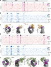
Fig. 4. Saturation mutagenesis on Fc corroborates the CU43i-FcE382S glycan structure.
a Heatmaps of deep mutational scanning results for IgG1 Fc mutants with (a) weaker and (b) tighter binding to CU43i. Average weaker/tighter binding scores per site are depicted as bar charts above the heatmaps, with sites highlighted if the score exceeds one (light blue/pink) or two (dark blue/magenta) standard deviations above the mean. c Average weaker (magenta/pink) and tighter (dark/light blue) binding scores, color-coded according to the threshold values described in (a–b), mapped onto Fc dimer structure with CU43i depicted in orange. N-glycan G0F glycan was docked in CU43i structure. d–e Heatmaps of deep mutational scanning results for IgG1 Fc mutants with (d) weaker and (e) tighter binding to EndoS2i. Average weaker/tighter binding scores per site are depicted as bar charts above the heatmaps, with sites highlighted if the score exceeds one (light blue/pink) or two (dark blue/magenta) standard deviations above the mean. f Average weaker (magenta/pink) and tighter (dark/light blue) binding scores, color-coded according to the threshold values described in (d, e), mapped onto Fc dimer structure with EndoS2i depicted in green. For statistical analysis of DMS data, we used two-sided Fisher’s exact test, followed by Bonferroni correction.
Fig. 5
Fig. 5. CU43 hydrolyzes IgG glycans by an inter-protomer mechanism.
a, b Mass spectrometry analysis of Fc WT CT containing N297- attached CT N-glycans incubated with (a) PBS or (b) after treatment with CU43 WT. Glycoforms on Fc are represented as cartoons. c-d) MS spectra of Fc WT glycoforms from Fc region of IgG1 homodimeric mutant variant harboring a A330W mutation in both protomers (chains A and B), incubated with (c) PBS or (d) after treatment with CU43 WT (1:1,000 enzyme:substrate molar ratio incubated during 3 hs at 37 °C). e, f MS spectra of IgG1 heterodimeric variant harboring a mutation A330W only in protomer A (chain A), incubated with (e) PBS or (f) after treatment with CU43 WT (1:1,000 enzyme:substrate molar ratio incubated during 3 h at 37 °C). g, h 5uM IgG1 heterodimeric variant harboring A330W only in protomer A (chain A) after treatment with 50 nM (g) CU43 or (h) EndoS2, and additionally treated with 75 nM IdeS peptidase and 50 nM BgaA (exo-galactosidase), after incubation for 1 h at 37 °C. In panels (E-H) the substrate was IgG Rituximab based KiH heterodimer: Protomer A: T366S/L368A/Y407V/A330W (−5Da); Protomer B: T366W ( + 85 Da). Assays were run in independent duplicates. Blue squares represent GlcNAc, red triangles represent Fucose, Green circles represent Mannose and yellow circles represent Galactose. Schematic Figures were created in BioRender. Sastre, D. (https://BioRender.com/uh09kf7). were created in (https://BioRender.com). Source data are provided as a Source Data file.
Fig. 6
Fig. 6. CU43 decouples the effector functions and neutralization capacities of IgG antibodies.
a FcγR humanized mice (n = 3/group) were administered I.v. with the indicated dose of the chimeric rat anti-mouse CD4 mAb (clone YTS191) expressed as afucosylated wild-type (WT) human IgG1. hFcyR mice, 6–8 weeks old. IVIG (IVIG Gammagard (Takeda) NDC 0944-2700-04) 5 mg/mouse (s.c.) + YTS191 afuc (aCD4) 10ug/mouse (i.v.) + CU43 1ug/mouse (i.v.); IVIG 5 mg/mouse (s.c.) + YTS191 afuc (aCD4) 10ug/mouse (i.v.) + PBS (i.v.); The levels of CD4 + T cells in the blood were determined by flow cytometry. Data are presented as mean values ± SEM from two independent experiments (n = 5 mice/group). b Schematic representation of CU43-Fc and Efgartigimod treatment to evaluate the capacity of neutralizing anti-influenza HA mAbs to confer in vivo protection against lethal influenza challenge in FcγR humanized mice. c–e FcγR humanized mice (n = 6-8 mice/group) were treated (i.v.) with the indicated dose of CU43-Fc or Efgartigimod (EFG) along with the neutralizing anti-HA mAb 7B2 (0.25 mg/kg, i.v.) one day prior to lethal challenge with H1N1 (Neth/09; i.n. 5mLD50). Weight (c, d) and survival (e) were monitored for a period of 14 days post-infection and compared between groups. AUC=area under the curve. Results are presented as the mean ± SEM from two independent experiments (n = 6–8 mice/group). f Schematic representation of the effect of IgG degrader vs IgG defeater on antibody-mediated functions and neutralization capacities. Schematics in panel 6b and 6f were created in BioRender. Sastre, D. (https://BioRender.com/uh09kf7)and Bournazos, S. (https://BioRender.com/7f9t1z4), respectively. Source data are provided as a Source Data file.
Fig. 7
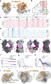
Fig. 7. CM49 utilizes a unique binding site on the Fc region to deglycosylate IgG antibodies.
a Two views of cartoon representation of AF3 models of CM49 (orange)-Fc dimer (gray) complex. N-glycan G0F was docked into AF-CM49 glycan binding pocket. b Neighbor-joining phylogenetic tree of corynebacterial and streptococcal ENGases with bootstrap values (1000 replicates). Protein sequence alignments were performed by MUSCLE and the phylogenetic tree was generated using MEGA v11. 0: indicates mechanism of multi-domain IgG-specific ENGases EndoS/S2; I: indicates mechanism of CU43 and its closest homologs; II: indicates the mechanism of CM49 and Csp50; ns, means non-IgG specific ENGase. c, d Heatmaps of deep mutational scanning results for IgG1 Fc mutants with (c) weaker and (d) tighter binding to CM49i. Average tighter/weaker binding scores per site are depicted as bar charts above the heatmaps, with sites highlighted if the score exceeds one (light blue/pink) or two (dark blue/magenta) standard deviations above the mean. e Average tighter (dark/light blue) and weaker (magenta/pink) binding scores, color-coded according to the threshold values described in (c, d), mapped onto Fc dimer structure (PDB code: 5JIH). For statistical analysis of DMS data we used two-sided Fisher’s exact test, followed by Bonferroni correction. e–i SAXS analysis of the CM49i-IgG1(Fc) complex. f SAXS scattering curve of CU49i, IgG1(Fc) and CU49i-IgG1(Fc). g P(r) functions distributions of CU49i, IgG1(Fc) and CU49i-IgG1(Fc) complex. G) Normalized Kratky plot CU49i, IgG1(Fc) and CU49i-IgG1(Fc) complex. h Ab initio modeling with GASBOR of the envelope of CM49i and superimposition of CM49i Alphafold structure prediction. i, j Ab initio modeling with GASBOR of the envelope of (i) CM49i-Fc and (j) superimposition of CM49i-Fc AF3 structure prediction.
Fig. 8
Fig. 8. CM49 binds a unique site on Fc IgG1 that does not overlap with any described hotspots for Fc binding.
Structural comparison of diversity of binding sites of proteins that specifically interact with IgG1 Fc. (1) top of CH2 binding: FcγRIIIA-Fc (PDB code: 1E4K), C1q-Fc (PDB code: 6FCZ), IdeS-Fc (PDB code: 8A47) and CU43i-Fc (PDB code: 8URO); (2) external surface of CH2-CH3 elbow: ProteinA-Fc (PDB code: 5U4Y), Protein G-Fc (PDB code: 1FCC), FcRn-Fc (PDB code: 7Q15), TRIM 21-Fc (PDB code: 2IWG) and EndoS-Fc (PDB code: 8A64); (3) CM49 bind to the inside surface of the CH2-CH3 elbow while no other Fc-binding protein or enzyme is known to bind to this site. Schematics were created in BioRender. Sastre, D. (https://BioRender.com/uh09kf7).

References

    1. Fairbanks, A. J. The ENGases: versatile biocatalysts for the production of homogeneous N-linked glycopeptides and glycoproteins. Chem. Soc. Rev.46, 5128–5146 (2017). - PubMed
    1. Trastoy, B. et al. Structural basis of mammalian high-mannose N-glycan processing by human gut Bacteroides. Nat. Commun.11, 899 (2020). - PMC - PubMed
    1. Sastre, D. E. et al. Human gut microbes express functionally distinct endoglycosidases to metabolize the same N-glycan substrate. Nat. Commun.15, 5123 (2024). - PMC - PubMed
    1. Seki, H. et al. Structural basis for the specific cleavage of core-fucosylated N-glycans by endo-beta-N-acetylglucosaminidase from the fungus Cordyceps militaris. J. Biol. Chem.294, 17143–17154 (2019). - PMC - PubMed
    1. Collin, M. & Olsen, A. EndoS, a novel secreted protein from Streptococcus pyogenes with endoglycosidase activity on human IgG. EMBO J.20, 3046–3055 (2001). - PMC - PubMed

MeSH terms

LinkOut - more resources