. 2025 Jul 3;11(1):304.
doi: 10.1038/s41420-025-02575-w.

FL3 mitigates cardiac ischemia-reperfusion injury by promoting mitochondrial fusion to restore calcium homeostasis

Affiliations

FL3 mitigates cardiac ischemia-reperfusion injury by promoting mitochondrial fusion to restore calcium homeostasis

Zikan Zhong et al. Cell Death Discov. .

Abstract

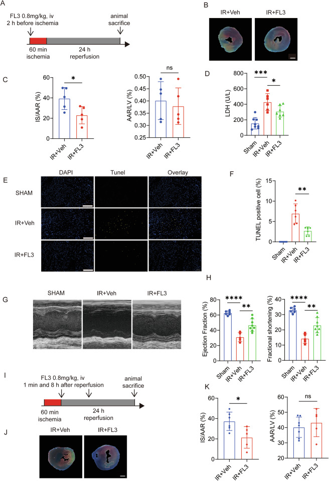
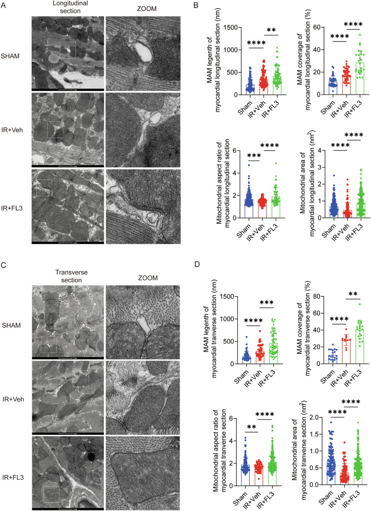
This study aims to investigate the therapeutic potential of Flavagline3 (FL3) in mitigating myocardial ischemia-reperfusion (IR) injury, with a specific focus on its regulatory effects on mitochondrial fusion, mitochondrial-endoplasmic reticulum (ER) interactions, and calcium homeostasis in cardiomyocytes. Using a well-established myocardial IR injury model in mice and primary cardiomyocytes treated with FL3, the study assessed its impact on mitochondrial dynamics and intracellular signaling processes. The results demonstrated that FL3 effectively reduced myocardial apoptosis, infarct size, and cardiac dysfunction caused by IR injury. Mechanistically, FL3 promoted mitochondrial fusion in a mitofusin1 (MFN1)-dependent manner, preserving mitochondrial function under stress conditions and enhancing cellular resilience. Furthermore, FL3 facilitated mitochondrial-ER crosstalk, which played a critical role in modulating intracellular calcium levels by optimizing the transfer of calcium ions between these two organelles. This balanced regulation of mitochondrial dynamics and calcium homeostasis was associated with improved survival and functionality of cardiomyocytes following IR injury. These findings suggest that FL3 exerts robust cardioprotective effects through its ability to promote mitochondrial fusion, enhance mitochondrial-ER interactions, and maintain calcium homeostasis. As a result, FL3 holds promise as a potential therapeutic agent for reducing myocardial damage and dysfunction associated with IR injury, offering valuable insights into novel approaches for cardioprotection.


Conflict of interest statement

Competing interests: The authors declare no competing interests. Ethics approvals: Animal experiments were approved by Shanghai General Hospital’s Ethics Committee. All procedures complied with ARRIVE reporting standards (see Supplementary File for checklist). No human subjects, tissues, or clinical data were involved in this study; therefore, informed consent and consent for publication of human images are not applicable.

Figures

Fig. 1
Fig. 1. Flavagline3 (FL3) Mitigates Myocardial Apoptosis Induced by Ischemia-Reperfusion (IR) Injury in the Heart.
A Experimental Design scheme: Mice were pretreated with a 0.8 mg/kg concentration of FL3 (IR + FL3 group) or DMSO (IR+Veh group) for two hours, and then both were subject to acute IR treatment (60 min ischemia followed by 24 hours reperfusion), all IR-treated animals were compared to the sham operation group. B Representative images of Evan’s blue and TTC double staining. C The quantitative data using images from panel B for infarct size (IF) (Left) and area at risk (AAR) (Right) (n = 5; scale bar: 1 mm). D The serum lactate dehydrogenase (LDH) concentration of sham operation group, IR+Veh group, and IR + FL3 group (n > 6). E, F The representative images (E) and statistical analysis data (F) of cardiac cell death indexed by TUNEL-positive cells (n = 8; scale bar: 0.2 mm). G, H Left ventricular ejection fraction (EF) and fractional shortening (FS) were evaluated by echocardiography (n = 6). I Experimental design scheme: Mice with acute IR injury (60 min ischemia followed by 24 h reperfusion) were divided into sham operation group, IR+Veh group, and IR + FL3 posttreatment group (administered after reperfusion onset). J Representative images of Evan’s blue and TTC double staining following posttreatment condition. K The quantitative data using images from panel J for infarct size (IF) (Left) and area at risk (AAR) (Right) (n = 5; scale bar: 1 mm). Data are presented as means ± SD. *P < 0.05, **P < 0.01, ***P < 0.001, ****P < 0.0001, ns, non-significant.
Fig. 2
Fig. 2. FL3 Attenuates Heart Failure and Cardiac Fibrosis Induced by IR Injury.
A Experimental design scheme: Mice were subjected to chronic IR injury (60 minutes ischemia followed by 4 weeks reperfusion). The animals were divided into three groups: sham operation group, IR+Veh group, and IR + FL3 treatment group (administered FL3 at 2 h before ischemia, followed by daily intraperitoneal injection of 0.8 mg/kg·d). B, C EF and FS were evaluated by echocardiography (n = 5 per group). D, E Myocardial infarction severity was assessed by Masson’s trichrome staining (Top) and HE staining (Bottom), as well as the heart weight to body weight ratio (HW/BW) and heart weight (n = 5 per group). These data were obtained from mice with chronic IR injury treated with either DMSO or FL3 (0.8 mg/kg·d), following the protocol outlined in A. F Mice subjected to chronic IR injury (60 min ischemia followed by 4 weeks reperfusion) were treated post-reperfusion with either DMSO (IR+Veh) or FL3 (administered at 1 minute and 4 h post-reperfusion, followed by daily intraperitoneal injections of 0.8 mg/kg·d). G, H EF and FS were measured by echocardiography (n = 6 [sham], n = 6 [IR+Veh group], and n = 7 [IR + FL3 group]). Data are presented as mean ± SD. *P < 0.05, **P < 0.01, ***P < 0.001.
Fig. 3
Fig. 3. FL3 Alleviates Hypoxia/Reoxygenation Induced Cardiomyocyte Apoptosis.
AC HL-1 cells were subjected to hypoxia/reoxygenation (HR) (12 h of hypoxia followed by 2 h of reoxygenation) with or without treatment with varying concentrations of FL3 (40 and 80 nM). Cleaved caspase-3 (H, n = 4) and cleaved PARP levels were evaluated using Western blots and statistical analysis. D, E The level of apoptosis was assessed by flow cytometry. F Representative images of neonatal rat ventricular myocytes (NRVMs) exposed to HR (24 h of hypoxia followed by 2 h of reoxygenation) with or without FL3 treatment (80 nM). G The proportion of TUNEL-positive cells in the culture medium was quantified. Data are expressed as mean ± SD. *P < 0.05, ****P < 0.0001.
Fig. 4
Fig. 4. FL3 Increases Mitochondria-Associated Endoplasmic Reticulum Membranes and Promotes Mitochondrial Fusion in the Myocardial IR Injury.
A Transmission electron microscopy (TEM) images (longitudinal section) of myocardial tissues from the sham operation, IR+Veh group, and IR + FL3 group were obtained to evaluate mitochondrial morphology. B Quantitative analysis of TEM images from A was performed, including measurements of mitochondria-associated endoplasmic reticulum membranes (MAM) length per mitochondrion (nanometers), percentage of endoplasmic reticulum coverage around mitochondria, mitochondrial aspect ratio, and mitochondrial area (nm²). C TEM images (transverse section) of myocardial tissues from the sham operation, IR+Veh group, and IR + FL3 group were obtained to evaluate mitochondrial morphology. D Quantitative analysis of TEM images from A was performed, including measurements of MAM length per mitochondrion (nanometers), percentage of endoplasmic reticulum coverage around mitochondria, mitochondrial aspect ratio, and mitochondrial area (nm²). Data are presented as means ± SD. **P < 0.01, ***P < 0.001, ****P < 0.0001.
Fig. 5
Fig. 5. FL3 Promotes Mitochondrial Fusion Through MFN1 During HR.
A Representative images of HL1 cells subjected to HR, stained with MitoTracker to assess mitochondrial fusion after FL3 treatment. B Quantitative parameters evaluated from the images, including mean form factor, average branch per mitochondrion, average branch length per mitochondrion (nanometers). C Representative images of NRVM cells under MFN1 and MFN2 knockdown conditions, stained with MitoTracker following FL3 treatment to assess mitochondrial fusion. D Quantitative parameters evaluated from C, including mean form factor, branches per mitochondrion, average branch length per mitochondrion (nanometers), and branch junctions per mitochondrion. E Representative images of NRVMs under MFN1 and MFN2 knockdown conditions, stained with MitoTracker after HR and FL3 treatment. F Quantitative parameters evaluated from (E), including mean form factor, branches per mitochondrion, average branch length per mitochondrion (nanometers), and branch junctions per mitochondrion. All quantitative data are presented as mean ± SD. *P < 0.05, **P < 0.01, ***P < 0.001, ****P < 0.0001, ns, non-significant.
Fig. 6
Fig. 6. FL3 Maintains Calcium Homeostasis and Protects Mitochondrial Function.
A Peak calcium ion flux in the mitochondrial matrix of NRVMs following histamine stimulation, FL3 treatment, and HR. B Mitochondrial matrix calcium ion concentrations in NRVMs under FL3 treatment and HR. CF Calcium ion concentrations in various subcellular compartments following FL3 treatment: (C) mitochondrial matrix, (D) MAM, (E) endoplasmic reticulum (ER), and (F) cytoplasm (Cyto). G, H JC-1 staining was used to assess mitochondrial membrane potential (n = 3), (G) Representative images, (H) Quantified JC-1 red/green fluorescence ratio. I, J Mitochondrial matrix calcium ion concentrations in NRVMs under FL3 treatment and HR analyzed with MFN1 knockdown (I), and with MFN2 knockdown (J), respectively. K, L Peak calcium ion flux in both the mitochondrial matrix and MAM under FL3 treatment in response to histamine stimulation: (K) mitochondrial matrix, (L) MAM. M The relative maximum rate of calcium release from the ER in HeLa cells following FL3 treatment in response to histamine stimulation. All quantitative data are presented as mean ± SD. NMX, normoxia; F/F0, peak-to-baseline ratio; *P < 0.05, **P < 0.01, ***P < 0.001, ****P < 0.0001, ns, non-significant.

References

    1. Hausenloy DJ, Yellon DM. Myocardial ischemia-reperfusion injury: a neglected therapeutic target. J Clin Invest. 2013;123:92–100. - PMC - PubMed
    1. Xiang Q, Yi X, Zhu XH, Wei X, Jiang DS. Regulated cell death in myocardial ischemia-reperfusion injury. Trends Endocrinol Metab. 2024;35:219–34. - PubMed
    1. Shen AC, Jennings RB. Kinetics of calcium accumulation in acute myocardial ischemic injury. Am J Pathol. 1972;67:441–52. - PMC - PubMed
    1. Kagan VE, Bayir HA, Belikova NA, Kapralov O, Tyurina YY, Tyurin VA, et al. Cytochrome c/cardiolipin relations in mitochondria: a kiss of death. Free Radic Biol Med. 2009;46:1439–53. - PMC - PubMed
    1. Weiss JN, Korge P, Honda HM, Ping P. Role of the mitochondrial permeability transition in myocardial disease. Circ Res. 2003;93:292–301. - PubMed

LinkOut - more resources