. 2025 Jul 4;16(1):6152.
doi: 10.1038/s41467-025-61440-7.

Discovery of a single-subunit oligosaccharyltransferase that enables glycosylation of full-length IgG antibodies in bacteria

Affiliations

Discovery of a single-subunit oligosaccharyltransferase that enables glycosylation of full-length IgG antibodies in bacteria

Belen Sotomayor et al. Nat Commun. .

Abstract

Human immunoglobulin G (IgG) antibodies are a major class of biotherapeutics and undergo N-linked glycosylation in their Fc domain, which is critical for immune functions and therapeutic activity. Hence, technologies for producing authentically glycosylated IgGs are in high demand. Previous attempts to engineer Escherichia coli for this purpose have met limited success due in part to the lack of oligosaccharyltransferase (OST) enzymes that can install N-glycans at the conserved N297 site in the Fc region. Here, we identify a single-subunit OST from Desulfovibrio marinus with relaxed substrate specificity that catalyzes glycosylation of native Fc acceptor sites. By chemoenzymatic remodeling the attached bacterial glycans to homogeneous, asialo complex-type G2 N-glycans, the E. coli-derived Fc binds human FcγRIIIa/CD16a, a key receptor for antibody-dependent cellular cytotoxicity (ADCC). Overall, the discovery of D. marinus OST provides previously unavailable biocatalytic capabilities and sets the stage for using E. coli to produce fully human antibodies.


Conflict of interest statement

Competing interests: M.P.D. and M.C.J. have financial interests in Gauntlet, Inc. and Resilience, Inc. M.P.D. also has financial interests in June Bio APS, Glycobia Inc., UbiquiTx Inc., and Versatope Therapeutics Inc. M.P.D.’s and M.C.J.’s interests are reviewed and managed by Cornell University and Stanford University, respectively, in accordance with their conflict-of-interest policies. M.P.D. and M.C.J. declare no additional competing interests. All other authors declare no competing interests.

Figures

Fig. 1
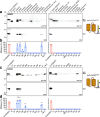
Fig. 1. Bioprospecting Desulfobacterota genomes for functional PglB homologs.
a Phylogenetic tree of the PglB homologs evaluated in this study. The curated list of enzymes was generated from a BLAST search using DaPglB and DgPglB as the query sequences. CjPglB and ClPglB were added for comparison. The tree was generated by the neighbor-joining method from multiple sequence alignment using Molecular Evolutionary Genetics Analysis version 11 (MEGA11) software. b Immunoblot analysis of periplasmic fractions from CLM24 cells transformed with plasmid pMW07-pglΔBCDEF encoding genes for biosynthesis of a modified C. jejuni heptasaccharide glycan (GalNAc5(Glc)GlcNAc), plasmid pBS-scFv13-R4DQNAT encoding the scFv13-R4DQNAT acceptor protein, and a derivative of plasmid pMLBAD encoding one of the PglB homologs as indicated. The first two lanes in left and right panels of (a) and (c) were loaded with the same positive and negative control samples (marked with asterisk). Blots were probed with polyhistidine epitope tag-specific antibody (anti-His) to detect the C-terminal 6x-His tag on the acceptor protein (top panel) and hR6 serum specific for the C. jejuni heptasaccharide glycan (bottom panel). Molecular weight (MW) markers are indicated on the left. The g0 and g1 arrows indicate un- and monoglycosylated acceptor proteins, respectively. Blots are representative of biological replicates (n = 3). Schematic created with BioRender. DeLisa, M. (2025) https://BioRender.com/wesqljn. c Glycosylation efficiency was determined by densitometric analysis, with data reported as mean ± SD. Red bars correspond to positive and negative controls generated with CjPglB and CjPglBmut, respectively; blue bars correspond to samples generated with Desulfovibrio PglBs. Statistical significance was determined by unpaired two-tailed Student’s t-test with calculated p-values represented as: *p < 0.05; ***p < 0.001; ns, not significant. Actual p-values relative to CjPglB are (from left-to-right): p = 0.541, p = 0.680, p = 0.0001, p = 0.0112, p = 0.0002, and p = 0.9586. Source data are provided as a Source Data file.
Fig. 2
Fig. 2. Glycosylation of non-canonical sequons by Desulfovibrio PglB homologs.
a Immunoblot analysis of periplasmic fractions from CLM24 cells transformed with the following plasmids: pMW07-pglΔBCDEF for making GalNAc5(Glc)GlcNAc; a derivative of pMLBAD encoding one of the PglB homologs as indicated; and pBS-scFv13-R4AQNAT encoding the scFv13-R4(N34L/N77L) acceptor protein with AQNAT sequon. Blots were probed with anti-His antibody (top panel) and hR6 serum (bottom panel). Molecular weight (MW) markers are indicated on the left. The g0 and g1 arrows indicate un- and monoglycosylated acceptor proteins, respectively. Blots are representative of biological replicates (n = 3). b Glycosylation efficiency was determined by densitometric analysis, with data reported as mean ± SD. Red bars correspond to positive and negative controls generated by CjPglB with scFv13-R4DQNAT or scFv13-R4(N34L/N77L)AQNAT as acceptors, respectively; blue bars correspond to samples generated with Desulfovibrio PglBs. (c, d) Same as (a, b) but with plasmid pBS-scFv13-R4QYNST encoding scFv13-R4(N34L/N77L) with QYNST sequon. Red bars correspond to positive and negative controls generated by CjPglB with scFv13-R4DQNAT or scFv13-R4(N34L/N77L)QYNST as acceptors, respectively; blue bars correspond to samples generated with Desulfovibrio PglBs. The first two lanes in left and right panels of (a) and (c) were loaded with the same positive and negative control samples (marked with asterisk). Schematics created with BioRender. DeLisa, M. (2025) https://BioRender.com/wesqljn. Statistical significance was determined by unpaired two-tailed Student’s t-test with calculated p-values represented as: *, p < 0.05; ***, p < 0.001; ns, not significant. Actual p-values relative to CjPglB (DQNAT) in panel (b) are (from left-to-right): p = 0.1072, p = 0.3838, p = 0.0008, p = 0.2436, and p = 0.2462. Actual p-values relative to CjPglB (DQNAT) in panel (d) are (from left-to-right): p = 0.6619, p = 0.0144, p = 0.0363 (DgPglB vs. DmPglB), p = 0.0017, and p = 0.0027. Source data are provided as a Source Data file.
Fig. 3
Fig. 3. Acceptor-site specificity of PglB homologs.
a Immunoblot analysis of periplasmic fractions from CLM24 cells transformed with the following plasmids: pMW07-pglΔBCDEF for making GalNAc5(Glc)GlcNAc; pMLBAD encoding DmPglB, DmPglBmut, CjPglB or CjPglBmut; and pBS-scFv13-R4XQNAT encoding the scFv13-R4 with each of the 20 amino acids in the –2 position of the C-terminal sequon as indicated. Blots were probed with anti-His antibody (top panel) and hR6 serum (bottom panel). Molecular weight (MW) markers are indicated on the left. The g0 and g1 arrows indicate un- and monoglycosylated acceptor proteins, respectively. Blots are representative of biological replicates (n = 3). Schematic created with BioRender. DeLisa, M. (2025) https://BioRender.com/wesqljn. b Heatmap analysis of the relative –2 amino acid preference of CjPglB, DgPglB, and DmPglB. Relative preferences (weaker = white; stronger = dark cyan) were determined based on densitometry analysis of the glycosylation efficiency for each acceptor protein in the anti-His immunoblot (see Supplementary Figs. 5, 6 for efficiency data). c Sequence logo showing experimentally determined acceptor-site specificity of DmPglB using glycoSNAP-based library screening of YebF(N24L)-Im7XXNXT. Source data are provided as a Source Data file.
Fig. 4
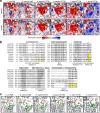
Fig. 4. Molecular determinants of relaxed acceptor-site specificity.
a Electrostatic potential of various OST peptide-binding pockets modeled with either DQNAT (top) or QYNST (bottom) acceptor peptides (yellow). Electrostatic surfaces were generated based on calculations using the adaptive Poisson-Boltzmann solver (APBS). b Sequence alignments of conserved, short motifs in eukaryotic STT3s (human and plant STT3A and STT3B, protozoan Leishmania major STT3D and Trypanosoma brucei TbSTTA) and bacterial ssOSTs (ClPglB, CjPglB, DgPglB, DmPglB, DiPglB). Alignments shown were made using Clustal Omega web server multiple alignment editor. Conserved residues are shaded gray while notable residues that deviate between eukaryotic and bacterial sequences are shaded yellow. c Structural model of QYNST peptide (yellow) in the peptide-binding pocket of the same OSTs in (a). Depicted in green are amino acids at the entrance to the peptide-binding cavity that cluster to create a positively charged patch in ClPglB but are neutral in all other OSTs. The SVXE/SVIE/TIXE motifs are depicted in gold.
Fig. 5
Fig. 5. Glycosylation of native acceptor site in IgG Fc domain by DmPglB.
a Non-reducing immunoblot analysis of protein A-purified proteins from whole-cell lysate of CLM24 cells transformed with the following plasmids: pMW07-pglΔBCDEF for making GalNAc5(Glc)GlcNAc (left) or pMW07-pglΔBICDEF for making GalNAc5GlcNAc (right); pMLBAD encoding CjPglB, DgPglB, DmPglB, or DmPglBmut; and pTrc99S-spDsbA-hinge-Fc encoding hinge-Fc derived from human IgG1. Blots were probed with anti-human IgG (anti-IgG) to detect human Fc (top panel) and hR6 serum (bottom panel). Molecular weight (MW) markers are indicated on the left. The g0, g1, and g2 arrows indicate un-, mono-, and diglycosylated Fc proteins, respectively. Blots are representative of biological replicates (n = 3). b Glycosylation efficiency was determined as above with data reported as the mean ± SD. Statistical significance was determined by unpaired two-tailed Student’s t-test with calculated p-values represented as: **p < 0.01; ***p < 001; ****p < 0.0001. Actual p-values in top panel are (from left-to-right): p < 0.0001 and p = 0.0002. Actual p-values in bottom panel are (from left-to-right): p = 0.0017 and p = 0.0017. c Same as in (a) but with JUDE-1 cells transformed with plasmid pMAZ360-YMF10-IgG encoding a full-length chimeric IgG1 specific for PA along with plasmids for glycan biosynthesis and ssOST as indicated. Asterisks denote band shifts due to glycosylation of HC-LC dimer. Schematics created with BioRender. DeLisa, M. (2025) https://BioRender.com/wesqljn. Source data are provided as a Source Data file.
Fig. 6
Fig. 6. Chemoenzymatic glycan remodeling of E. coli-derived hinge-Fc proteins.
a Schematic representation of the chemoenzymatic reaction for trimming and remodeling hinge-Fc glycans. b Immunoblot analysis of the four E. coli-derived glycoforms (from left to right): aglycosylated hinge-Fc, glycosylated GalNAc5GlcNAc-hinge-Fc, GlcNAc-hinge-Fc, and G2-hinge-Fc. Blot was probed with anti-human IgG (anti-IgG) to detect human Fc. Molecular weight (MW) markers are indicated on the left. The g0, g1, and g2 arrows indicate un-, mono-, and diglycosylated Fc proteins, respectively. Blot is representative of biological replicates (n = 3). c ELISA analysis of same constructs in (b) with FcγRIIIA-V158 as immobilized antigen. Data are average of biological replicates (n = 3) ± SD. Schematics created with BioRender. DeLisa, M. (2025) https://BioRender.com/wesqljn.

Update of

Similar articles

Cited by

References

    1. Abu-Qarn, M., Eichler, J. & Sharon, N. Not just for Eukarya anymore: protein glycosylation in bacteria and archaea. Curr. Opin. Struct. Biol.18, 544–550 (2008). - PubMed
    1. Apweiler, R., Hermjakob, H. & Sharon, N. On the frequency of protein glycosylation, as deduced from analysis of the SWISS-PROT database. Biochim. Biophys. Acta1473, 4–8 (1999). - PubMed
    1. Stanley, P., Moremen, K. W., Lewis, N. E., Taniguchi, N. & Aebi, M. In Essentials of Glycobiology, Edn. 4th. (eds. Varki, A. et al.) 103−116 (Cold Spring Harbor (NY), 2022).
    1. Khoury, G. A., Baliban, R. C. & Floudas, C. A. Proteome-wide post-translational modification statistics: frequency analysis and curation of the swiss-prot database. Sci. Rep.1, 90 (2011). - PMC - PubMed
    1. Seeberger, P.H., Freedberg, D.I. & Cummings, R.D. In Essentials of Glycobiology, 4th Edn. (eds. Varki, A. et al.) 771−784 (Cold Spring Harbor (NY), 2022).

MeSH terms

LinkOut - more resources