. 2025 Jul 3;16(1):489.
doi: 10.1038/s41419-025-07791-x.

The dual mechanism of m6A demethylase ALKBH5 in regulating energy metabolism during exposure to MC-LR

Affiliations

The dual mechanism of m6A demethylase ALKBH5 in regulating energy metabolism during exposure to MC-LR

Xiaoya Sun et al. Cell Death Dis. .

Abstract

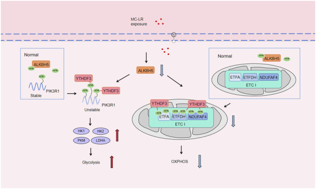
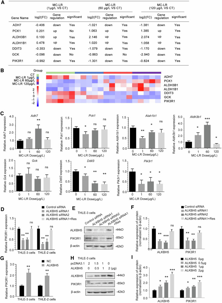
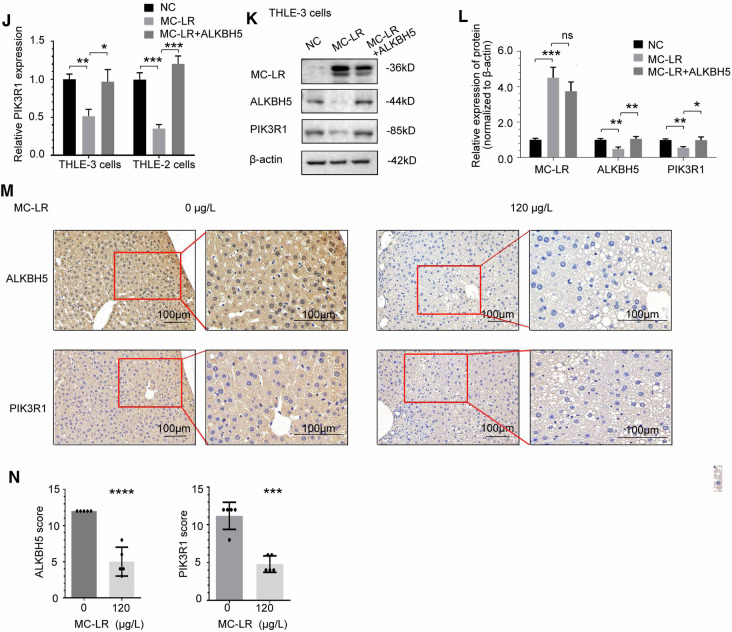
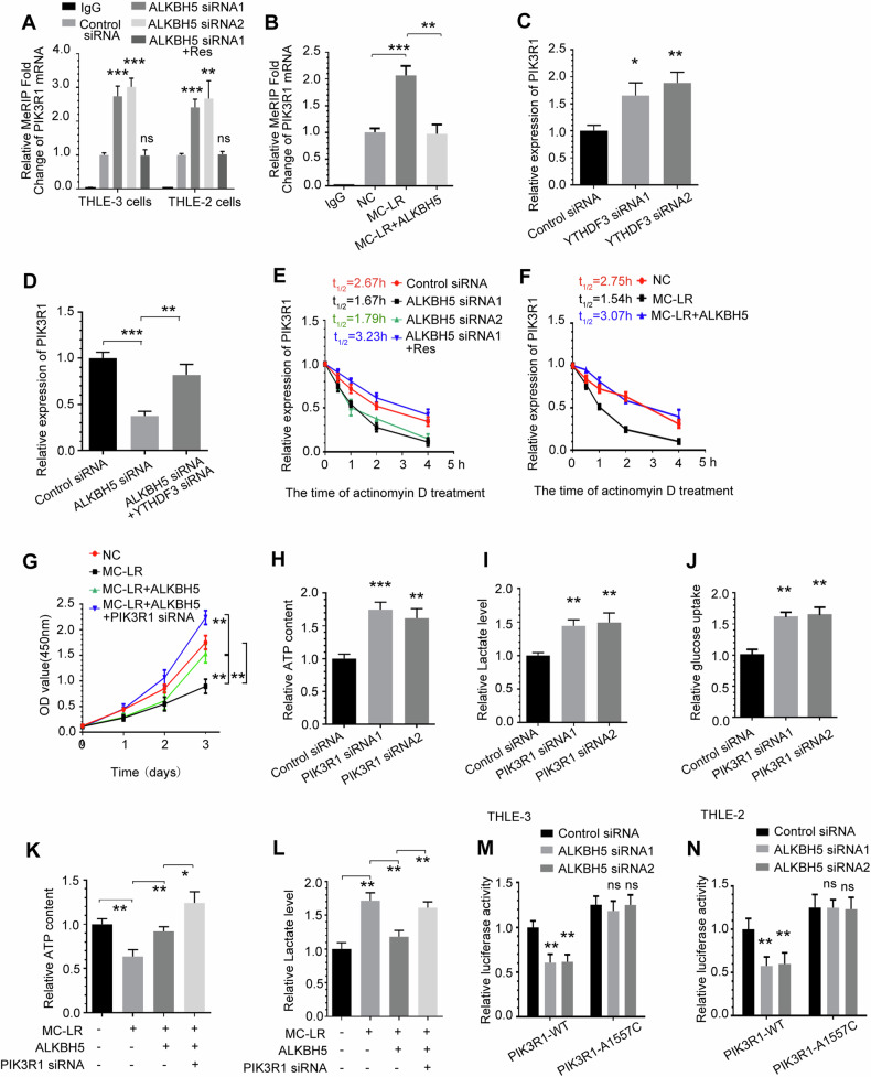
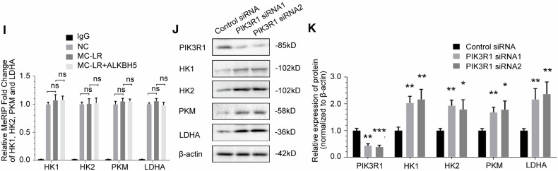
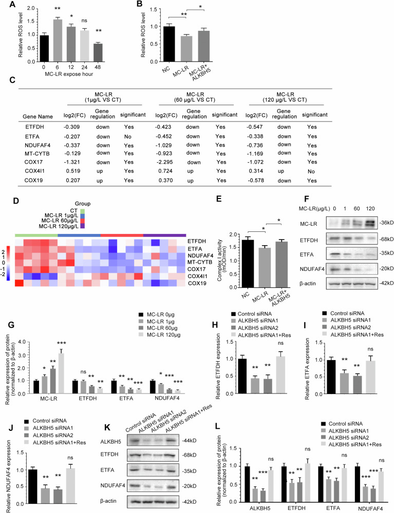
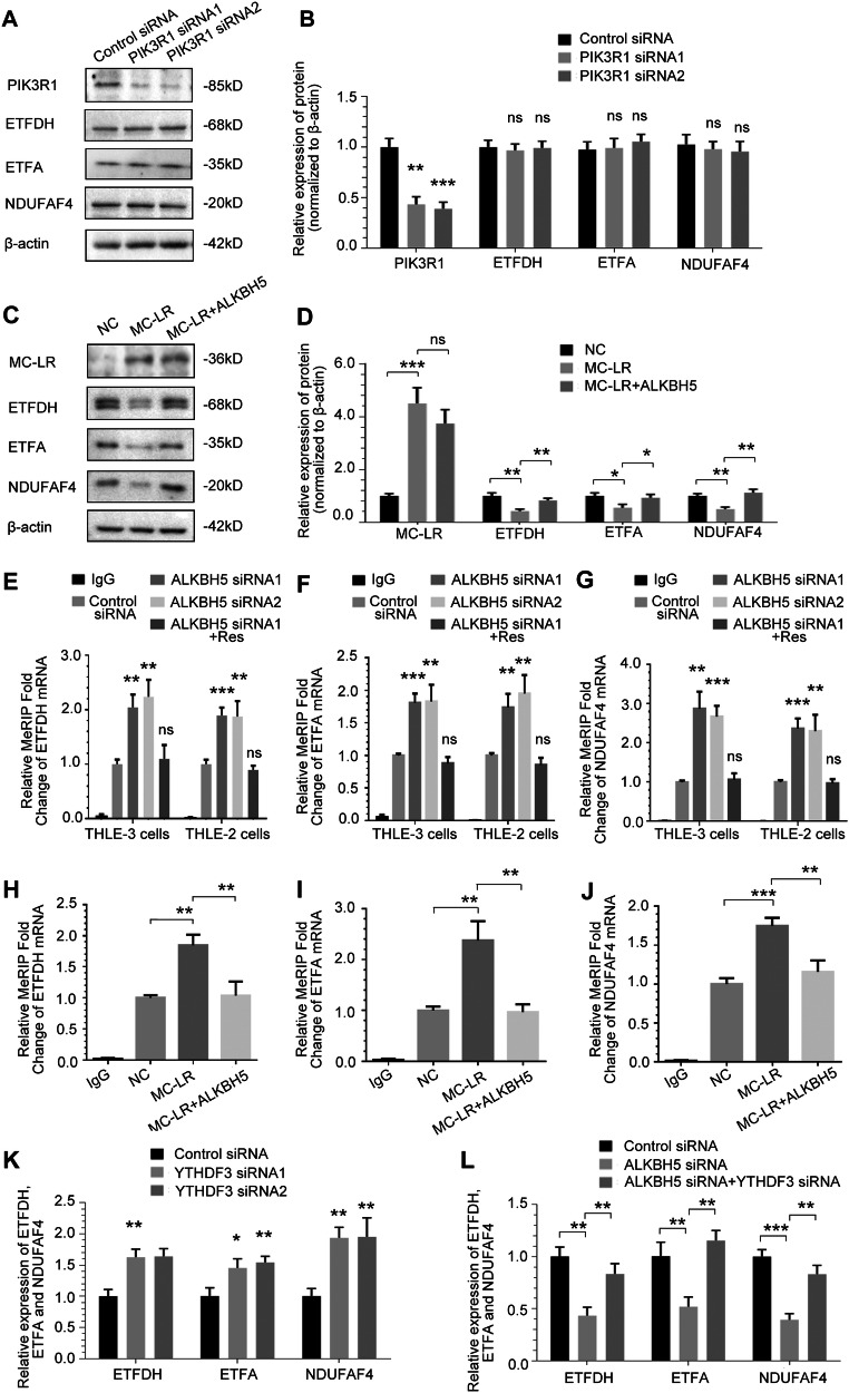
Exposure to MC-LR has been shown to cause multiple organ injury, particularly liver injury, and altered energy metabolism is closely linked. As an effective and efficient way to regulate biological gene expression, N(6)-methyladenosine(m6A) modification plays an important role in liver injury caused by microcystin-LR(MC-LR) exposure. For the first time, we reveal the dual mechanism by which AlkB homolog 5(ALKBH5) regulates energy metabolism through an m6A-YTHDF3-dependent mechanism. After MC-LR exposure, low levels of ALKBH5 increased the m6A modification of Phosphoinositide-3-Kinase Regulatory Subunit 1(PIK3R1) and m6A methylation was located at A1557. PIK3R1-m6A was recognised by YTH N6-Methyladenosine RNA Binding Protein F3(YTHDF3), which reduced the stability of PIK3R1 RNA, thereby inhibiting PIK3R1 expression and ultimately promoting glycolysis. In concert, low-level ALKBH5 inhibit oxidative phosphorylation by down-regulating the expression of Electron Transfer Flavoprotein Dehydrogenase(ETFDH), Electron Transfer Flavoprotein Subunit Alpha(ETFA) and NADH:Ubiquinone Oxidoreductase Complex Assembly Factor 4(NDUFAF4) through an m6A-YTHDF3-dependent mechanism. This dual mechanism has been shown to adversely affect cell survival in MC-LR exposed environments by significantly reducing ATP levels. This study reveals for the first time the signalling pathway and molecular mechanism of MC-LR exposure to liver injury through ALKBH5-mediated m6A modification, providing new protective and therapeutic principles.Subject terms: m6A modification; Oxidative phosphorylation; Glycolysis The mechanism of m6A demethylase ALKBH5 in regulating energy metabolism during exposure to MC-LR. Created with BioRender.com.


Conflict of interest statement

Competing interests: The authors declare no competing interests.

Figures

None
The mechanism of m6A demethylase ALKBH5 in regulating energy metabolism during exposure to MC-LR. Created with BioRender.com.
Fig. 1
Fig. 1. MC-LR exposure induces inhibition of hepatocyte growth and increases RNA m6A methylation.
A Pathological observation of liver tissue from MC-LR exposed mice (HE staining). B Effects of MC-LR exposure on proliferation of THLE-3 cells. C Effects of MC-LR exposure on colony forming ability in THLE-3 cells. The m6A methylation level of the liver tissues of mice with exposure to MC-LR was detected by m6A dot blot assay (D) and corresponding quantification (E). The m6A methylation level of total RNA in THLE-3 after 48 h of different doses of MC-LR was detected by m6A dot blot assay (F) and corresponding quantification(G). Data are means ± SD from three independent experiments. *P ≤ 0.05; **P ≤ 0.01; ***P ≤ 0.001; ****P ≤ 0.0001.
Fig. 2
Fig. 2. ALKBH5 expression is repressed and mediates upregulation of m6A modification induced by MC-LR.
A In RNA-seq data analysis, m6A writers, erasers, and readers in liver tissues from MC-LR exposed mice compared to vehicle control mice. B Heatmap of RNA expression of the m6A writer, eraser and reader genes. C RT-PCR measurements of the enzymes and mediators for RNA m6A methylation in liver tissues from MC-LR exposed mice. D ALKBH5 protein expression decreased in liver tissue of mice exposed to MC-LR. RT-PCR (E) and Western blotting measurements (F) of ALKBH5 expression in human liver cells with 5 μM MC-LR exposure. Effect of overexpressing exogenous ALKBH5 vectors on the change in m6A level with MC-LR exposure was detected by m6A dot blot assay (G) and corresponding quantification (H). Effect of overexpressing exogenous METTL3 (I and J) and METTL14 (K and L) vectors on the change in m6A level with MC-LR exposure was detected by m6A dot blot assay and corresponding quantification. Data are means ± SD from three independent experiments. *P ≤ 0.05; **P ≤ 0.01; ***P ≤ 0.001.
Fig. 3
Fig. 3. ALKBH5 mediates MC-LR exposure-induced altered energy metabolism and cell proliferation inhibition.
Intracellular ATP levels (A) and extracellular lactate production (B) in liver tissue of mice exposed to MC-LR compared to vehicle control mice. The effect of ALKBH5 knockdown on intracellular ATP levels (C), extracellular lactate production (D), glucose uptake (E) and NAD+ /NADH ratios (F) was measured compared to control cells. G, H Effects of knockdown ALKBH5 on the colony-forming abilities of THLE-3 cells. I, J Effect of overexpression of ALKBH5 on colony forming ability of THLE-3 cells. Effects of ALKBH5 on intracellular ATP levels (K), extracellular lactate production (L), glucose uptake (M) and NAD+ /NADH ratios (N) of THLE-3 cells following MC-LR exposure. Effect of ALKBH5 overexpression (O and P) or knockdown (Q and R) on the colony forming ability of THLE-3 cells after exposure to MC-LR. Data are means ± SD from three independent experiments. *P ≤ 0.05; **P ≤ 0.01; ***P ≤ 0.001.
Fig. 4
Fig. 4. MC-LR exposure inhibits PIK3R1 expression mediated by ALKBH5.
A Cell proliferation-related genes in liver tissue of mice exposed to MC-LR in RNA-seq data analysis. B Heatmap of RNA expression of the cell proliferation-related genes. C RT-PCR measurements of the related genes in liver tissue from MC-LR exposed mice. ALKBH5 knockdown reduced PIK3R1 mRNA (D) and protein (E, F) expression. Overexpression of ALKBH5 increased PIK3R1 mRNA (G) and protein (H, I) expression. MC-LR exposure resulted in the downregulation of PIK3R1 mRNA (J) and protein (K, L) expression, which was rescued by overexpression of ALKBH5. M Representative images of immunohistochemistry (IHC) analysis of ALKBH5 and PIK3R1 in liver tissue from MC-LR exposed mice. N IHC staining scores of ALKBH5 and PIK3R1 in liver tissue of mice exposed to MC-LR. Data are means ± SD from three independent experiments. *P ≤ 0.05; **P ≤ 0.01; ***P ≤ 0.001; ****P ≤ 0.0001.
Fig. 5
Fig. 5. ALKBH5 enhances PIK3R1 RNA stability in an m6A-YTHDF3-dependent manner and inhibits glycolysis.
A PIK3R1 mRNA m6A methylation was increased upon knockdown of ALKBH5. B PIK3R1 mRNA m6A methylation was increased upon MC-LR exposure, which could be rescued by overexpressing ALKBH5. C YTHDF3 knockdown increased PIK3R1 mRNA expression. D YTHDF3 knockdown can reverse the change in PIK3R1 mRNA expression induced by ALKBH5 knockdown. E The half-life of PIK3R1 mRNA was shortened by ALKBH5 knockdown in THLE-3 cells. F The half-life of PIK3R1 mRNA was shortened upon MC-LR exposure, which could be rescued by overexpression of ALKBH5. G Effects of overexpression of ALKBH5 or simultaneous knockdown of PIK3R1 on cell proliferation inhibition induced by MC-LR exposure. Intracellular ATP levels (H), extracellular lactate production (I) and glucose uptake (J) were measured in PIK3R1 knockdown cells compared to control cells. Effects of ALKBH5 overexpression or PIK3R1 knockdown on intracellular ATP levels (K) and extracellular lactate production (L) in cells exposed to MC-LR. In THLE-3 (M) and THLE-2 (N) cells, ALKBH5 knockdown reduced the activity of PIK3R1’s WT. The A1557C mutation of PIK3R1 resulted in increased activity and was not affected by ALKBH5 knockdown. Data are means ± SD from three independent experiments. *P ≤ 0.05; **P ≤ 0.01; ***P ≤ 0.001.
Fig. 6
Fig. 6. ALKBH5 inhibits glycolytic pathway enzymes through the mediation of PIK3R1.
A RNA-seq data analysis of glycolytic pathway enzyme genes in liver tissue from MC-LR exposed mice. B Heatmap of RNA expression of the glycolytic pathway enzyme genes. C, D The effect of ALKBH5 knockdown on the expression of PIK3R1 and glycolytic pathway enzymes in THLE-3 cells, as detected by Western blotting and quantified. E, F The effect of MC-LR exposure on the expression of MC-LR, ALKBH5, PIK3R1 and glycolytic pathway enzymes in mouse liver tissues, as detected by Western blotting and quantified. G, H The effect of overexpression of ALKBH5 on the expression levels of MC-LR, ALKBH5, PIK3R1 and glycolytic pathway enzymes proteins in THLE-3 cells treated with MC-LR, as detected by Western blotting and quantified. I m6A modifications of HK1, HK2, PKM and LDHA were not affected by MC-LR exposure and ALKBH5 overexpression. J, K The effect of PIK3R1 knockdown on the expression of glycolytic pathway enzymes in THLE-3 cells, as detected by Western blotting and quantified. Data are means ± SD from three independent experiments. *P ≤ 0.05; **P ≤ 0.01; ***P ≤ 0.001.
Fig. 7
Fig. 7. Depresses of ALKBH5 suppresses mitochondrial oxidative phosphorylation during MC-LR exposure.
A Cellular ROS levels determined in THLE-3 cells cultured under MC-LR exposure for 0, 6, 12, 24, 48 h. B Effects of overexpression of ALKBH5 on Cellular ROS levels exposed to MC-LR. C Analysis of mitochondrial oxidative phosphorylation enzyme genes in liver tissue of mouse exposed to MC-LR in RNA-seq data analysis. D Heatmap of RNA expression of the mitochondrial oxidative phosphorylation enzyme genes. E Specific regulation of ETC complex I activity by overexpression of ALKBH5 under MC-LR exposure. F, G The effect of MC-LR exposure on the expression of ETFDH, ETFA and NDUFAF4 in mouse liver tissue, as detected by Western blotting and quantified. Effect of ALKBH5 knockdown on ETFDH, ETFA and NDUFAF4 RNA (H–J) and protein (K, L) expression in THLE-3 cells. Data are means ± SD from three independent experiments. *P ≤ 0.05; **P ≤ 0.01; ***P ≤ 0.001.
Fig. 8
Fig. 8. ETFDH, ETFA and NDUFAF4 mRNAs are targets of ALKBH5 demethylation.
A, B The effect of PIK3R1 knockdown on the expression of ETFDH, ETFA and NDUFAF4 in THLE-3 cells, as detected by Western blotting and quantified. C, D The effect of overexpression of ALKBH5 on the expression levels of ETFDH, ETFA and NDUFAF4 proteins in THLE-3 cells treated with MC-LR,as detected by Western blotting and quantified. EG ETFDH, ETFA and NDUFAF4 m6A modifications were increased upon knockdown of ALKBH5. HJ ETFDH, ETFA and NDUFAF4 RNA m6A modifications was increased upon MC-LR exposure, which could be rescued by overexpressing ALKBH5. K YTHDF3 knockdown increased ETFDH, ETFA and NDUFAF4 mRNA expression. L YTHDF3 knockdown can reverse the change in ETFDH, ETFA and NDUFAF4 mRNA expression induced by ALKBH5 knockdown. Data are means ± SD from three independent experiments. *P ≤ 0.05; **P ≤ 0.01; ***P ≤ 0.001.

Similar articles

References

    1. Yang F, Huang F, Feng H, Wei J, Massey IY, Liang G, et al. A complete route for biodegradation of potentially carcinogenic cyanotoxin microcystin-LR in a novel indigenous bacterium. Water Res. 2020;174:115638. 10.1016/j.watres.2020.115638. - PubMed
    1. Buratti FM, Manganelli M, Vichi S, Stefanelli M, Scardala S, Testai E, et al. Cyanotoxins: producing organisms, occurrence, toxicity, mechanism of action and human health toxicological risk evaluation. Arch Toxicol. 2017;91:1049–130. 10.1007/s00204-016-1913-6. - PubMed
    1. Merel S, Walker D, Chicana R, Snyder S, Baures E, Thomas O. State of knowledge and concerns on cyanobacterial blooms and cyanotoxins. Environ Int. 2013;59:303–27. 10.1016/j.envint.2013.06.013. - PubMed
    1. Mugani R, El KF, Redouane EM, Haida M, Aba RP, Essadki Y, et al. Unlocking the potential of bacterioplankton-mediated microcystin degradation and removal: A bibliometric analysis of sustainable water treatment strategies. Water Res. 2024;255:121497. 10.1016/j.watres.2024.121497. - PubMed
    1. Li X, Li L, Huang Y, Wu H, Sheng S, Jiang X, et al. Upstream nitrogen availability determines the Microcystis salt tolerance and influences microcystins release in brackish water. Water Res. 2024;252:121213. 10.1016/j.watres.2024.121213. - PubMed

MeSH terms

LinkOut - more resources