

Green synthesis of silver nanoparticles using Magnolia alba leaf extracts and evaluating their antimicrobial, anticancer, antioxidant, and photocatalytic properties
- PMID: 40610522
- PMCID: PMC12229685
- DOI: 10.1038/s41598-025-08468-3
Green synthesis of silver nanoparticles using Magnolia alba leaf extracts and evaluating their antimicrobial, anticancer, antioxidant, and photocatalytic properties
Abstract
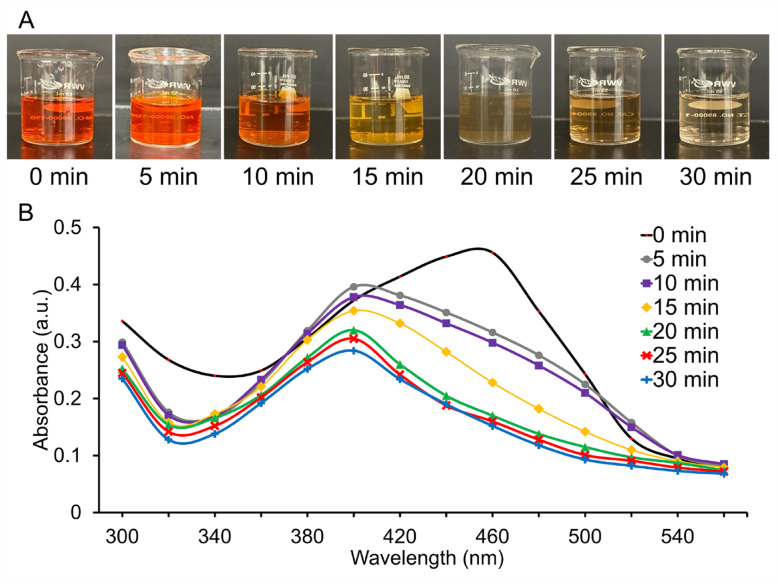
The biosynthesis of silver nanoparticles has recently emerged as a promising approach in nanomedicine, particularly for targeted therapeutic applications. Green synthesized (plant-based) nanoparticles have been shown to offer enhanced reduction efficiency, greater bioavailability, and improved stability compared to synthetic nanoparticles. Here, we report the green synthesis of silver nanoparticles (AgNPs) using Magnolia alba leaf extract (MLE). The formation of these Magnolia-derived silver nanoparticles (MAgNPs) was verified through UV-Vis spectroscopy with a surface plasmon resonance peak at 440 nm, and further characterized by scanning electron microscopy, which showed that the MAgNPs have a mean diameter of 40 nm and a spherical morphology. MAgNPs exhibited significant antibacterial activity against Escherichia coli, Klebsiella pneumoniae, Pseudomonas aeruginosa, Enterococcus faecalis, methicillin-resistant and -sensitive Staphylococcus aureus, with a minimum inhibitory concentration of 0.00043 mg/mL and a minimum bactericidal concentration of 0.00043 mg/mL and 0.0017 mg/mL, respectively. Disc diffusion and plaque assays with MAgNPs demonstrated strong antifungal activity against Candida albicans, with a zone of inhibition of 14 mm, and antiviral activity against T7 bacteriophage (p = 0.0004). In vitro studies with HCT-116 human colon cancer cells, MAgNPs exhibited bi-phasic, dose-dependent inhibition of viability with a 20-40% reduction, surpassing the positive control Camptothecin. Antioxidant assays indicated that MAgNPs showed significantly higher antioxidant activity compared to MLE, with enhanced Total Flavonoid Content (p = 0.0066), Total Phenol Content (p = 0.0013), and Total Antioxidant Capacity (p = 0.0051). Additionally, MAgNPs showed efficient photocatalytic degradation of the azo bond in methyl orange within 30 min. To our knowledge, this is the first report on the biosynthesis of MAgNPs and their multifunctional properties, highlighting the promise of MAgNPs in biomedical and environmental fields. (Insert attached Graphical abstract).
Keywords: Antimicrobial; Cancer; Candida; HCT-116; MRSA; Pneumoniae; T7 Bacteriophage.
© 2025. The Author(s).
Conflict of interest statement
Declarations. Competing interest: The authors declare no competing interests. Ethical approval: This research was conducted within the institutional guidelines of both The Master’s University and Biola University. The Magnolia alba is not listed on Convention on International Trade in Endangered Species of Wild Fauna and Flora (CITES) or any endangered species list. This research did not harm or harass any endangered plant species and complied with the Plant Protection Act (PPA) and International Plant Protection Convention (IPPC) regulations. As the research was conducted within California, it adhered to relevant state and federal regulations. Furthermore, the Nagoya Protocol on Access and Benefit-Sharing is not applicable to this study.
Figures


Similar articles
-
Biosynthesis and characterization of silver nanoparticles from Asplenium dalhousiae and their potential biological properties.PLoS One. 2025 Jun 30;20(6):e0325533. doi: 10.1371/journal.pone.0325533. eCollection 2025. PLoS One. 2025. PMID: 40587502 Free PMC article.
-
Green synthesis of silver nanoparticles (AgNPs) from G. stearothermophilus GF16: stable and versatile nanomaterials with antioxidant, antimicrobial, and catalytic properties.Microb Cell Fact. 2025 Aug 19;24(1):189. doi: 10.1186/s12934-025-02815-9. Microb Cell Fact. 2025. PMID: 40830875 Free PMC article.
-
Green synthesis of silver nanoparticles from plant Astragalus fasciculifolius Bioss and evaluating cytotoxic effects on MCF7 human breast cancer cells.Sci Rep. 2025 Jul 15;15(1):25474. doi: 10.1038/s41598-025-05224-5. Sci Rep. 2025. PMID: 40664749 Free PMC article.
-
Green Synthesis of Titanium Dioxide Nanoparticles: Physicochemical Characterization and Applications: A Review.Int J Mol Sci. 2025 Jun 6;26(12):5454. doi: 10.3390/ijms26125454. Int J Mol Sci. 2025. PMID: 40564917 Free PMC article. Review.
-
Green Synthesis of Silver Nanoparticles Using Plant Extracts: A Comprehensive Review of Physicochemical Properties and Multifunctional Applications.Int J Mol Sci. 2025 Jun 27;26(13):6222. doi: 10.3390/ijms26136222. Int J Mol Sci. 2025. PMID: 40650001 Free PMC article. Review.
References
-
- Wu, Q., Miao, W.-S., Zhang, Y.-D., Gao, H.-J. & Hui, D. Mechanical properties of nanomaterials: A review. Nanotechnol. Rev.9(1), 259–273 (2020).
-
- Rajeshkumar, S. & Bharath, L. V. Mechanism of plant-mediated synthesis of silver nanoparticles - A review on biomolecules involved, characterisation and antibacterial activity. Chem. Biol. Interact.273, 219–227 (2017). - PubMed
MeSH terms
Substances
LinkOut - more resources
Full Text Sources
Medical
Miscellaneous
