. 2025 Jul 3;8(1):987.
doi: 10.1038/s42003-025-08382-4.

Potential role of developmental experience in the emergence of the parvo-magno distinction

Affiliations

Potential role of developmental experience in the emergence of the parvo-magno distinction

Marin Vogelsang et al. Commun Biol. .

Abstract

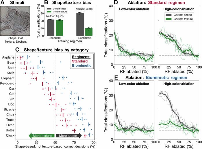
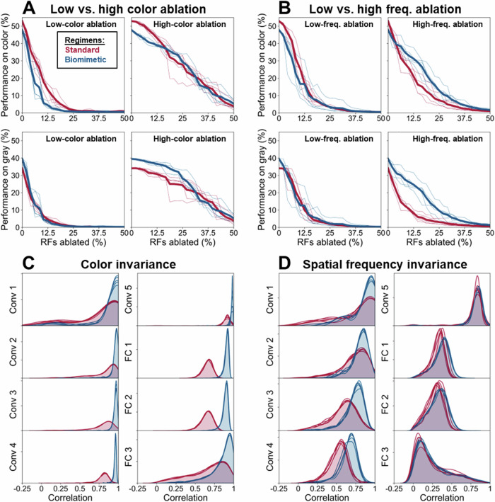
While the division of the early visual pathway into parvo- and magnocellular systems with distinct response properties has long been established as a prominent organizing principle in mammalian visual systems, the factors that lead to its emergence remain unclear. Here, we provide a potential account of this emergence based on early sensory development. Specifically, we propose that the temporal confluence in the developmental progression of spatial frequency and chromatic sensitivities may significantly shape corresponding neuronal response properties characteristic of this division. Receptive field analyses of deep networks trained on developmentally inspired 'biomimetic' protocols support this proposal in both the spatial and temporal domain. Further, biomimetic training induces a more human-like bias towards global shape processing, potentially driven by magnocellular units. These results have implications for the emergence of a key aspect of visual pathway organization and applied relevance for the design of training procedures for computational vision systems.


Conflict of interest statement

Competing interests: The authors declare no competing interests.

Figures

Fig. 1
Fig. 1. Results of receptive field analysis.
First-layer RFs following training with the standard (A) and biomimetic (B) regimen. Color (C) and spatial frequency (D) distributions of individual RFs of both models. Scatter plots depicting the joint frequency and color coding of individual RFs following training with the standard (E) and biomimetic regimen (F). Depicted here are results obtained with the first training run; qualitatively similar outcomes of five training runs with different random initializations are shown in Supplementary Fig. 2.
Fig. 2
Fig. 2. Results of texture/shape analysis.
A Exemplar shape/texture cue conflict stimulus, reprinted from Geirhos et al., depicting the local texture of an elephant and the global shape of a cat. B Percentage of total classifications correct in terms of shape, correct in terms of texture, or neither (i.e., incorrect). Error bars represent the standard error across the five different training runs with random initializations, and dots depict results of each individual run. C Percentage of shape-based correct classifications, as opposed to texture-based correct classifications, for each of the 16 different super-classes. Classifications that were inconsistent with both texture and shape classes are not included in this computation. Shown here are results of the five individual training runs (dots), along with their means (bars). Depicted on the y-axis are shape categories. Shape/texture bias as a function of the proportion of the ablated least color-sensitive (left) and most color-sensitive (right) units, for the standard (D) and biomimetic (E) regimen. Depicted are the five training runs (thin lines) as well as their means (thick lines). The dashed line, plotted at 1/16, indicates the chance classification level expected of a network generating random responses.
Fig. 3
Fig. 3. Additional characterization of training regimens.
Classification performance on color (top) and grayscale (bottom) images when ablating the least vs. most color-sensitive (A) as well as least vs. most high spatial frequency tuned (B) first-layer receptive fields. Depicted are results of five training runs with different random initializations (thin lines), along with their means (thick lines). As there are 1000 ImageNet classes, the chance level is 0.1%. Distribution of correlations of neural activations across layers between full-color and grayscale images (C) and between full-frequency and blurred images (D). Depicted are superimposed results of the five individual network training runs.
Fig. 4
Fig. 4. Results of simulations with 3D-CNNs.
Histograms of color (A) and spatial frequency (B) metrics for receptive fields of the biomimetic and standard model when training is conducted with video inputs to a 3D-CNN. C Relationship between temporal RF properties (using the temporal variation metric; see Methods) and spatial RF characteristics (using the color and spatial frequency metrics akin to those used before) of the biomimetic model. The ellipse marks the five receptive fields exhibiting the greatest temporal variation. These RFs also show consistently low color and spatial frequency tuning. D Visualization of the five most temporally varied receptive fields of the biomimetic model. E Depiction of biphasic receptive field, adapted from the neurophysiological study by De Valois et al.. F Relationship between the temporal variation metric and spatial RF characteristics (color and spatial frequency metrics) of the standard model. The ellipses mark the five receptive fields exhibiting the greatest temporal variation. G Visualization of the five most temporally varied receptive fields of the standard model. All plots in Fig. 4 have been generated based on the first out of five training runs with different random initializations. Results of all five training runs are shown in Supplementary Fig. 19.

References

    1. Livingstone, M. & Hubel, D. Segregation of form, color, movement, and depth: anatomy, physiology, and perception. Science240, 740–749 (1988). - PubMed
    1. Shapley, R. Visual sensitivity and parallel retinocortical channels. Annu. Rev. Psychol.41, 635–658 (1990). - PubMed
    1. Shapley, R. Parallel retinocortical channels: X and Y and P and M. In Advances in psychology(Vol. 86, pp. 3-36). North-Holland (1992).
    1. Wiesel, T. N. & Hubel, D. H. Spatial and chromatic interactions in the lateral geniculate body of the rhesus monkey. J. Neurophysiol.29, 1115–1156 (1966). - PubMed
    1. Schiller, P. H. & Malpeli, J. G. Functional specificity of lateral geniculate nucleus laminae of the rhesus monkey. J. Neurophysiol.41, 788–797 (1978). - PubMed

LinkOut - more resources