. 2025 Jul 3;15(1):23811.
doi: 10.1038/s41598-025-09733-1.

Felodipine re-positioned as a neuroprotectant via improved optic nerve head blood circulation in retinal ischemic rabbits and ocular hypertensive rats

Affiliations

Felodipine re-positioned as a neuroprotectant via improved optic nerve head blood circulation in retinal ischemic rabbits and ocular hypertensive rats

Takazumi Taniguchi et al. Sci Rep. .

Abstract

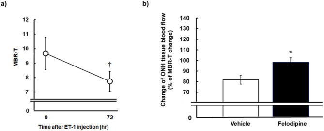
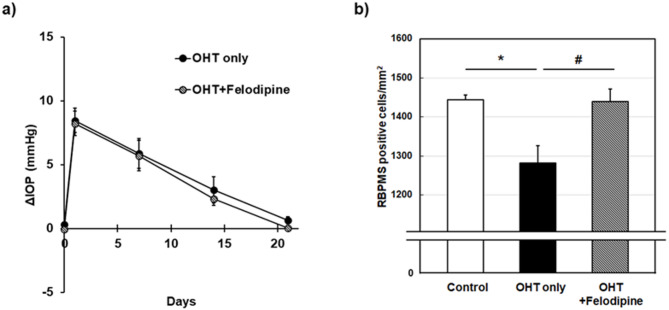
The vasodilatory and neuroprotective properties of felodipine were characterized in normal and ischemic rabbit eyes, an ocular hypertension (OHT) rat model, and cultured human neuron-like cells. Blood flow in the optic nerve head (ONH) was measured using laser speckle flowgraphy following intravitreally injected felodipine into normal and ischemic rabbit eyes receiving endothelin-1 injection. Felodipine concentrations in the retina-choroidal tissue were determined using mass spectrometry, and ocular safety was assessed using in-life examinations and histopathology in rabbits. The number of retinal ganglion cells (RGCs) was counted following intravitreal felodipine injection in hypertonic saline-induced OHT rats. The in vitro neuroprotective effects of felodipine against vincristine-induced nuclear loss and neurite shortening were evaluated in differentiated SH-SY5Y neuron-like cells. Felodipine increased ONH blood flow in a dose-dependent manner in normal rabbit eyes. High felodipine dosage (780 nmol/eye) improved blood circulation in endothelin-1-induced ischemic eyes. The ONH blood flow response to felodipine correlated with tissue concentration-time profiles. Even high dose of felodipine did not cause any ocular toxicity. Furthermore, felodipine protected SH-SY5Y cells against vincristine-induced damage and prevented OHT-induced RGC cell loss following its administration (40 nmol/eye). Felodipine can act as a neuroprotectant against glaucomatous neuropathy via improved ONH microcirculation and direct neuroprotection of RGCs.

Keywords: Calcium channel blocker; Felodipine; Glaucoma; Neuroprotection; Ocular blood flow; Retinal ganglion cells.


Conflict of interest statement

Declarations. Competing interests: The authors declare no competing interests.

Figures

Fig. 1
Fig. 1
Effects of intravitreal injection of felodipine on tissue blood flow (MBR-T) in the optic nerve head (ONH) in conscious rabbits. Vehicle or felodipine (7.8, 78, and 780 nmol/eye) was injected into the vitreous of the left eye of each animal, whereas the right eye remained untreated. ONH tissue blood flow was determined using laser speckle flowgraphy before injection, and 24 and 72 h after injection. ONH blood flow was expressed as a percentage of the pretreatment value (baseline). Each value represents the mean ± SEM of 1 to 4 eyes. Statistically significant differences are shown as * P < 0.05, ** P < 0.01, compared to the vehicle group at the respective timepoints (Dunnett’s multiple comparison test).
Fig. 2
Fig. 2
Preventive effects of intravitreal injection of felodipine against impaired optic nerve head (ONH) blood flow induced by endothelin-1 (ET-1) in the rabbit eyes. Under a conscious condition, each animal received intravitreal injection of vehicle or 780 nmol/eye felodipine prior to ET-1 (5 pmol/eye) intravitreal injection. ONH tissue blood flow was measured by laser speckle flowgraphy before and 72 h after ET-1 injection. (a) Effect of ET-1 alone on tissue blood flow (MBR-T) measured before and 72 h after concurrent injection of ET-1 and vehicle. P < 0.05 (Paired t-test) compared with the baseline (time 0). (b) Effect of vehicle or felodipine on ET-1-induced ONH blood flow impairment. ONH blood flow measured at 72 h after ET-1 injection is expressed as a percentage of the pre-treatment value (baseline) in each group. *P < 0.05 (Student’s t-test) compared with vehicle. Each value represents the mean ± SEM of 7 to 8 eyes.
Fig. 3
Fig. 3
Tissue felodipine concentration-time profiles in the retina-choroid of the rabbit eyes receiving intravitreal injection of felodipine (7.8, 78, and 780 nmol/eye). Eyes were enucleated 4, 24, and 72 h following its injection. The retina-choroid were isolated and tissue felodipine concentrations were determined using mass spectrometry. Each value represents the mean ± SEM (n = 4–6 eyes).
Fig. 4
Fig. 4
Neuroprotective effects of felodipine against vincristine-induced cellular damage in differentiated SH-SY5Y cells. The nucleus and neurites were stained with the cell membrane stain kit and hoechst33342 following treatment with vehicle (control, 0.1% DMSO), vincristine alone (200 nM), or felodipine (0.1 and 0.3 µM) plus vincristine. (a) Representative images of nuclear (blue) and neurite (orange) staining with or without drug treatments. The scale bar shows 500 μm. (b,c) Quantitative data to assess neuroprotective effects of felodipine. The number of nucleus and total neurite length per number of the nucleus were quantified in each analyzed field. Each value represents the mean ± SEM of 3 wells. * P < 0.05 *** P < 0.001, compared with the control group by either Student’s t-test or Aspin-Welch test. # P < 0.05, ### P < 0.001, compared with vincristine alone by Dunnett’s multiple comparison test.
Fig. 5
Fig. 5
Protective effects of intravitreal injection of felodipine against ocular hypertension (OHT)-induced retinal ganglion cell damage in rats. (a) OHT was induced by episcleral venous injection of hypertonic saline into the left eye of each animal and the contralateral eye remained untreated. Felodipine (40 nmol/eye) was intravitreally injected twice on the day of OHT surgery and 11 days later. The IOP profiles were presented as the difference between the OHT eyes and the contralateral eyes. Each value represents the mean ± SEM of 5 eyes. (b) Determination of retinal ganglion cells (RGCs) density based on the staining of RNA-binding protein with multiple splicing (RBPMS) in OHT rats. Eyes were enucleated from normal (control), OHT without drug treatment (OHT only) and OHT rats receiving intravitreally injected felodipine (OHT + felodipine) three weeks following OHT surgery. RGCs were detected by immunostaining using an anti-RBPMS antibody on whole-mounted retinas. Each value represents the mean ± SEM of 5–6 eyes. * P < 0.05, compared with the control group by Aspin-Welch test; # P < 0.05, compared with the OHT only group by Student’s t-test.

References

    1. Weinreb, R. N. et al. Primary open-angle glaucoma. Nat. Rev. Dis. Primers 2 Preprint at (2016). 10.1038/nrdp.2016.67 - PubMed
    1. Tham, Y. C. et al. Global prevalence of glaucoma and projections of glaucoma burden through 2040: A systematic review and meta-analysis. Ophthalmology121, 2081–2090 (2014). - PubMed
    1. Sharif, N. A. Elevated intraocular pressure and glaucomatous optic neuropathy: genes to disease mechanisms, therapeutic drugs, and gene therapies. Pharmaceuticals16, 870 (2023). - PMC - PubMed
    1. Tribble, J. R. et al. Neuroprotection in glaucoma: mechanisms beyond intraocular pressure Lowering. Mol. Asp Med.92, Preprintathttpsdoiorg101016jmam2023101193 (2023). - PubMed
    1. He, S., Stankowska, D. L., Ellis, D. Z., Krishnamoorthy, R. R. & Yorio, T. Targets of neuroprotection in glaucoma. J. Ocul Pharmacol. Ther.34, 85–106 (2018). - PMC - PubMed

MeSH terms

LinkOut - more resources