. 2025 Jul 3;24(1):268.
doi: 10.1186/s12933-025-02772-y.

Single nuclei RNA-sequencing unveils alveolar macrophages as drivers of endothelial damage in obese HFpEF-related pulmonary hypertension

Affiliations

Single nuclei RNA-sequencing unveils alveolar macrophages as drivers of endothelial damage in obese HFpEF-related pulmonary hypertension

Era Gorica et al. Cardiovasc Diabetol. .

Abstract

Background: Pulmonary hypertension (PH) is a frequent complication in obese patients showing heart failure with preserved ejection fraction (HFpEF) and correlates with poor prognosis. PH associated with cardiometabolic HFpEF (PH-cHFpEF) is characterized by inflammation and metabolic dysregulation. Alterations in the immune landscape, particularly activation of alveolar macrophages (AMs), may propagate the inflammatory response and lead to endothelial damage and vascular remodeling in the lung. Whether AMs contribute to PH in cardiometabolic HFpEF remains elusive.

Purpose: The present study investigates the role of alveolar macrophages in PH-cHFpEF.

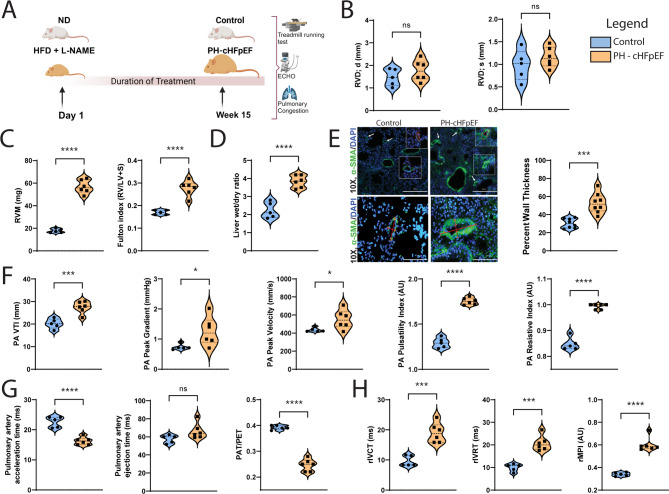
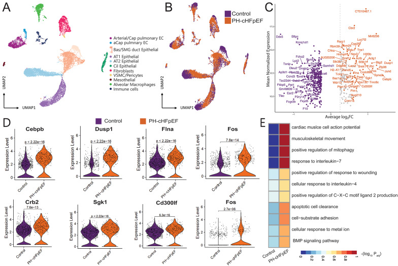
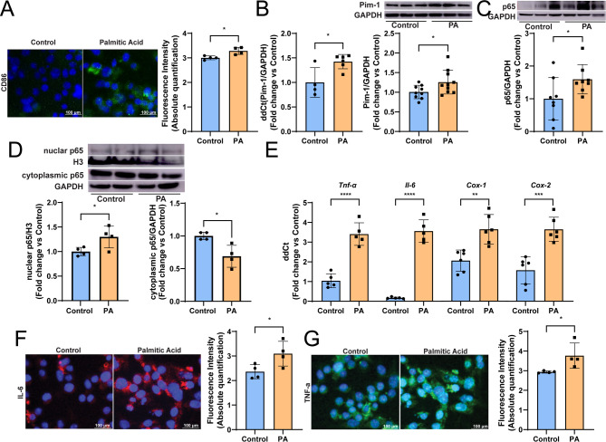
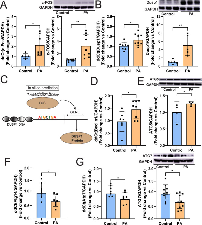
Methods: Mice subjected to high-fat diet and L-NAME treatment for 15 weeks were used as experimental model of PH-cHFpEF. At the end of the treatment, echocardiography and treadmill exhaustion tests were performed. Single nucleus RNA-sequencing (snRNA-seq) was employed to study the AMs transcriptional landscape and cell-cell interactions. In vitro experiments were performed to study the mechanisms underlying metabolic stress-induced macrophage dysfunction using palmitic acid (PA), co-culture experiments were used to investigate the crosstalk between macrophages and endothelial cells.

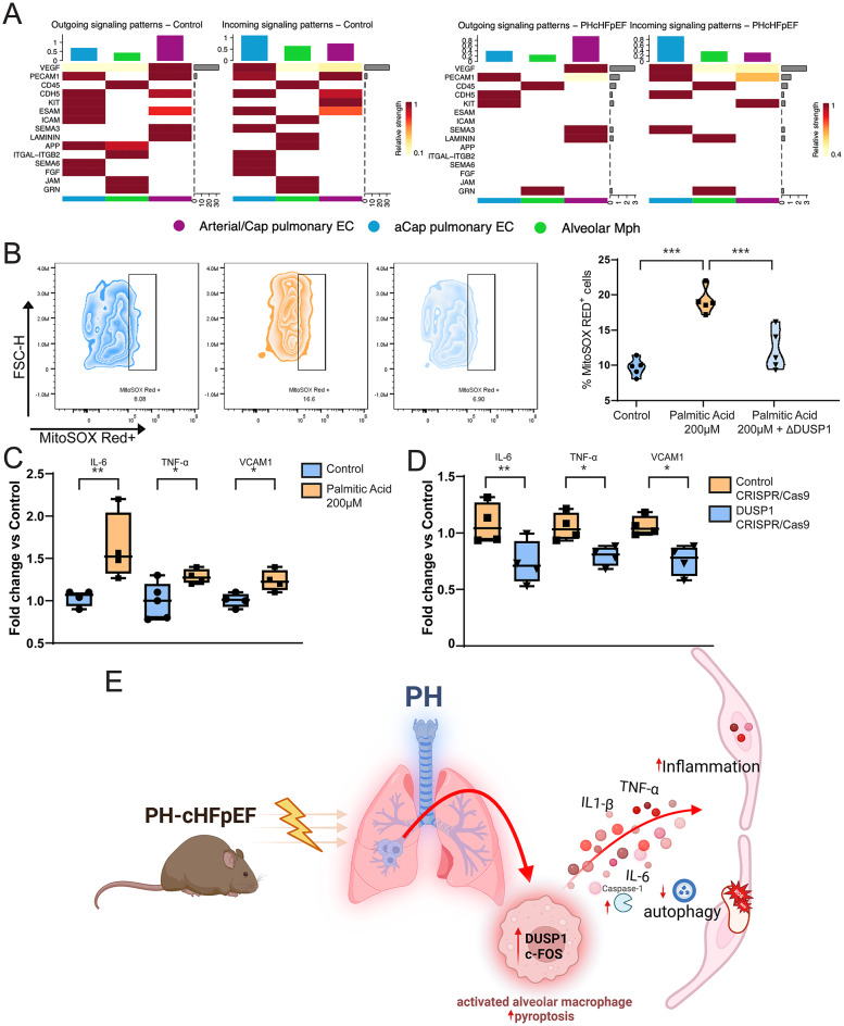
Results: Compared with control mice, PH-cHFpEF animals displayed right ventricular dysfunction, vascular remodeling and increased pulmonary pressure. SnRNA-seq of mouse lungs revealed transcriptional alterations in AMs, with a significant reduction in their abundance in PH-cHFpEF mice. These changes were associated with dysregulation of transcriptional programs involved in pyroptosis, defective autophagy and inflammation in AMs from PH-cHFpEF vs. control mice, as shown by the upregulation of c-Fos, Dusp1, Pim-1 and Ccn1. STRING analysis revealed a molecular link between these partners and highlighted c-Fos/Dusp-1 as a central axis of AMs cell death and inflammation. Metabolic stress induced by PA in isolated murine macrophages recapitulated c-Fos/Dusp-1 activation as well as IL-1β, TNF-α, and Caspase-1 upregulation resulting in inflammation, impaired autophagy and enhanced pyroptosis. Moreover, c-Fos/Dusp1 activation in macrophages promoted secretion of pro-inflammatory chemokines leading to endothelial dysfunction in a paracrine manner. Dusp1 knockdown rescued autophagy and pyroptosis while mitigating macrophage-driven inflammation and endothelial damage.

Conclusions: PH-cHFpEF is characterized by AMs activation, upregulation of the cFos/Dusp-1 pathway and subsequent pyroptosis and inflammation in alveolar macrophages. Our findings highlight the role of AMs as putative targets for preventing endothelial damage in experimental PH-cHFpEF.

Keywords: Alveolar macrophages; Endothelial damage; Inflammation; PH; cHFpEF.


Conflict of interest statement

Declarations. Competing interests: Era Gorica and Francesco Paneni are Guest Editors of the collection “Cardiometabolic HFpEF with focus on type 2 diabetes mellitus”, but haven’t been involved in handling this manuscript during the submission or review process.The authors declare no competing interests.

Figures

Fig. 1
Fig. 1
Figure 1. Right ventricular dysfunction and pulmonary hypertension in a mouse model of cardiometabolic disease. A Schematic showing the experimental mouse model of PH-cHFpEF. B Right ventricular diastolic diameter (RVD; d) and right ventricular systolic diameter (RVD; s) in control and PH-cHFpEF mice. C-D Right ventricular remodeling and function, assessed by RV mass, Fulton Index and Liver wet/dry ratio in PH-cHFpEF as compared to control mice. E Representative α-SMA immunofluorescence staining of lung tissue sections from mice (left), example of determining the wall thickness of a pulmonary vessel (right), and the respective quantification of the medial wall thickness. α-smooth muscle actin (α-SMA, GREEN); 4′,6-diamidino-2-phenylindole (DAPI, BLUE). F Pulmonary artery velocity time integral (PA VTI), pulmonary artery peak gradient (PA Peak Gradient), pulmonary artery peak velocity (PA Peak Velocity), pulmonary artery pulsatility index (PA Pulsatility Index), and pulmonary artery resistive index (PA Resistive Index). G Pulmonary artery acceleration time, pulmonary artery ejection time, pulmonary acceleration time to ejection time ratio (PAT/PET) in control and PH-cHFpEF mice. H Right ventricular contraction time (rVCT), right ventricular relaxation time (rVRT), and right ventricular myocardial performance index (rMPI). Data are presented as mean ± SD on a sample size of at least n=5. Statistical analyses were performed using either one-way ANOVA with Bonferroni post hoc testing or Student's t-test, as appropriate. A p-value ≤0.05 was considered significant.
Fig. 2
Fig. 2
Molecular phenotyping of the HFpEF lung by snRNAseq. UMAP representation of pulmonary cells colored by A cell type and B condition (control vs. PH-cHFpEF). C Volcano plot highlighting the top 90 most upregulated genes in murine alveolar macrophages in control (purple) and PH-cHFpEF (orange) mice. D Violin plots depicting the normalized expression levels of PH-cHFpEF-specific genes in alveolar macrophages, supported by mean comparison p-values via the non parametric Wilcoxon Rank Sum statistical test. E Heatmap presenting the most enriched gene ontology (GO) biological processes pathways of alveolar macrophages in PH-cHFpEF mice, computed as Normalized (-(log10 Padj)). Cap: Capillary, aCap EC: aerocytes Capillary endothelial cells, Bas/ SMG duct: Basal submucosal gland duct, AT: alveolar type/ pneumocytes, Cil: Ciliated airway/respiratory cells, VSMC: Vascular smooth muscle cells.
Fig. 3
Fig. 3
Dusp1 involvement in inflammation, autophagy and pyroptosis in PH-cHFpEF mice. A Protein–protein interactions from the STRING database show the top-ranking proteins interacting with DUSP1. B Molecular analysis of mouse lung specimens of control and PH-cHFpEF mice. C ELISA assay in lung tissues of control vs. PH-cHFpEF mice. D Olink proteomics of mouse serum. Data are presented as mean ± SD on a sample size of at least n = 4. Statistical analyses were performed using Student’s t-test, as appropriate. A p-value ≤ 0.05 was considered significant.
Fig. 4
Fig. 4
Metabolic stress and inflammation in RAW 264.7 murine macrophages in vitro. A Immunofluorescence staining for macrophage polarization. B qPCR and WB of Pim-1 in control and PA-treated murine macrophages. C WB of total p65 expression and D WB of nuclear and cytosolic fractions of p65. E qPCR analysis of inflammatory markers expression in murine macrophages. F-G Immunofluorescence staining and the respective fluorescence intensity quantification of IL-6 and TNF-α in PA-treated cells. Data are presented as mean ± SD on a sample size of at least n = 4. Statistical analyses were performed using Student’s t-test. A p-value ≤ 0.05 was considered significant.
Fig. 5
Fig. 5
Macrophage pyroptosis and oxidative stress in RAW 264.7 murine macrophages. A qPCR and WB of BAX/Bcl2 ratio in murine macrophages treated with PA. B Flow cytometry analysis of Annexin V and Propidium Iodide (PI). C qPCR and ELISA assay of Caspase-1. D WB of CCN1 protein in control and PA-treated macrophages. E qPCR and ELISA assay of IL-1β in murine macrophages. E Immunofluorescence staining of DHE oxidative stress marker. Data are presented as mean ± SD on a sample size of at least n = 5. Statistical analyses were performed using Student’s t-test. A p-value ≤ 0.05 was considered significant.
Fig. 6
Fig. 6
Macrophage cFos and DUSP1 activation and autophagy dysregulation in RAW 267.4. A-B PCR and WB analysis of cFOS and DUSP1 in control and PA-treated murine macrophages. C Bioinformatic tools predicting potential binding sites of cFOS on the DUSP1 gene promoter. D-E qPCR of Beclin1 and WB of Atg5 in control and PA-treated macrophages. F qPCR of Atg14 in control vs. PA-treated cells. G) qPCR and WB of Atg7 in murine macrophages treated either with control or PA. Data are presented as mean ± SD on a sample size of at least n = 5. Statistical analyses were performed using Student’s t-test. A p-value ≤ 0.05 was considered significant.
Fig. 7
Fig. 7
Effect of DUSP1 targeted via CRISPR/Cas9 on pyroptosis and autophagy of RAW 264.7 murine macrophages exposed to metabolic stress. A Cell viability analysis in control CRISPR/Cas9 and DUSP1 CRISPR/Cas9 knockout cells exposed to PA treatment. B WB of p65 and C CCN1 comparing DUSP1 CRISPR/Cas9 targeted vs. control CRISPR/Cas9 targeted cells exposed to PA. D qPCR of inflammatory and E pyroptotic genes in DUSP1 CRISPR/Cas9 targeted murine macrophages ad exposed to metabolic stress. F WB of ATG7 comparing DUSP CRISPR/Cas9 targeted vs. control CRISPR/Cas9 targeted cells exposed to PA. Data are presented as mean ± SD from at least three independent experiments. Statistical analyses were performed using Student’s t-test. A p-value ≤ 0.05 was considered significant.
Fig. 8
Fig. 8
Macrophage conditioned media and endothelial cell activation in vitro. A Heatmap-based bar plots depicting the outgoing and incoming signaling exchanged among alveolar macrophages and endothelial cells control and PH-cHFpEF mice. B MitoSOX Red Flow Cytometry of human aortic endothelial cells (HAECs) treated in control, PA-treated, and ΔDUSP1 PA-treated RAW 264.7 macrophage conditioned media. C-D qPCR of pro-inflammatory cytokines IL-6 and TNF-α, as well as the adhesion molecule VCAM-1 in endothelial cells exposed control, PA-treated, and ΔDUSP1 PA-treated macrophage conditional media. E Representative scheme of crosstalk between alveolar macrophages and endothelial cells in PH. Data are presented as mean ± SD on a sample size of at least n = 3. Statistical analyses were performed using Student’s t-test. A p-value ≤ 0.05 was considered significant.

References

    1. Min J, Feng R, Badesch D, Berman-Rosenzweig E, Burger C, Chakinala M, et al. Obesity in pulmonary arterial hypertension (PAH): the pulmonary hypertension association registry (PHAR). Ann Am Thorac Soc. 2020;18(2):229–37. - PMC - PubMed
    1. Moles VM, Grafton G. Pulmonary hypertension in heart failure with preserved ejection fraction. Cardiol Clin. 2022;40(4):533–40. - PubMed
    1. Guazzi M. Pulmonary hypertension in heart failure preserved ejection fraction: prevalence, pathophysiology, and clinical perspectives. Circ Heart Fail. 2014;7(2):367–77. - PubMed
    1. Mair KM, Gaw R, MacLean MR. Obesity, estrogens and adipose tissue dysfunction—implications for pulmonary arterial hypertension. Pulm Circ. 2020;10(3):2045894020952019. - PMC - PubMed
    1. Humbert M, Kovacs G, Hoeper MM, Badagliacca R, Berger RMF, Brida M, et al. 2022 ESC/ERS guidelines for the diagnosis and treatment of pulmonary hypertension. Eur Heart J. 2022;43(38):3618–731. - PubMed

MeSH terms

Substances

LinkOut - more resources