[Preprint]. 2025 Jun 20:2025.06.18.660432.
doi: 10.1101/2025.06.18.660432.

Complete cross strain protection against congenital cytomegalovirus infection requires a vaccine encoding key antibody (gB) and T-cell (immediate early 1 protein) viral antigens

Affiliations

Complete cross strain protection against congenital cytomegalovirus infection requires a vaccine encoding key antibody (gB) and T-cell (immediate early 1 protein) viral antigens

K Yeon Choi et al. bioRxiv. .

Abstract

Background: Cytomegalovirus is a leading cause of congenital disease and multiple strains enable congenital CMV (cCMV) from both primary and non-primary infection. A cross-strain protective cCMV vaccine is a high priority. The guinea pig is the only small animal model for cCMV and guinea pig cytomegalovirus (GPCMV) encodes functional homolog proteins including cell entry gB glycoprotein and non-structural immediate early 1 protein (IE1), essential for lytic infection. A gB vaccine antibody response fails to provide horizontal protection against highly cell-associated clinical GPCMV strain TAMYC compared to prototype strain 22122. Previously, a recombinant defective adenovirus (Ad) vaccine encoding IE1, a T-cell antigen, provided high-level cCMV protection. In this study, we hypothesized that a combined Ad-based strategy encoding trimeric gB complex and IE1 (AdgB+AdIE1) could improve cross-strain protection against cCMV compared to a gB vaccine (AdgB).

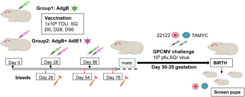
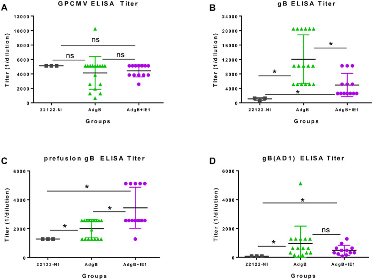
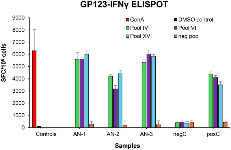
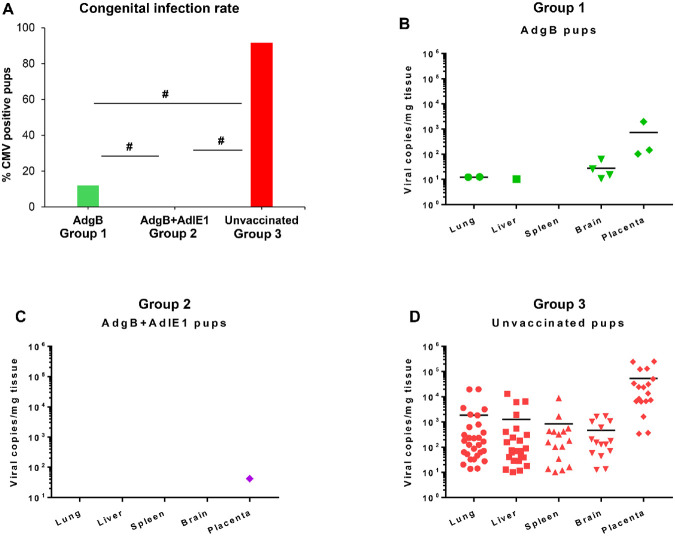
Methods: A preconception vaccine study evaluated the immune response and ability of vaccines to provide cross-strain protection against cCMV. Seronegative female animals were assigned into three vaccine groups: Group 1 (AdgB); Group 2 (AdgB+AdIE1); Group 3 (no vaccine). Animals were vaccinated following a previously defined protocol and antibody ELISAs were used to evaluate gB immune response (AD1, prefusion gB and wild type gB). Additionally, an IFNγ-ELISPOT assay evaluated IE1 T-cell response. During second trimester dams were challenged with GPCMV (22122 and TAMYC) and pregnancy went to term where viral loads in pup target organs and placentas were evaluated.

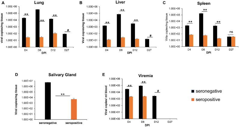
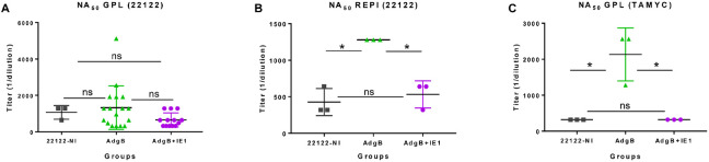
Results: Vaccinated dams elicited a higher neutralizing antibody response to gB than natural convalescent immunity and antibodies recognized homolog AD1 gB domain as well as prefusion gB with response surpassing natural immunity. Group 2 animals additionally elicited a T-cell response to IE1. Evaluation of viral load in pups demonstrated that AdgB+AdIE1 vaccine reduced GPCMV transmission to below detectable limits compared to 91.7% in unvaccinated group. In contrast, AdgB reduced cCMV transmission to 12% in pups.

Conclusion: Complete cross-strain cCMV protection is a significant milestone in this model and achieved by inclusion of an antibody response to trimeric gB and T-cell response to IE1. Importantly, gB and IE1 responses can synergize and increase protection against cCMV unlike prior approaches.

Keywords: CMV vaccine; IE1; congenital CMV; cytomegalovirus; gB; guinea pig; placenta.


Conflict of interest statement

Conflict of Interest The authors declare that the research was conducted in the absence of any commercial or financial relationships that could be construed as a potential conflict of interest.

Figures

Figure 1.
Figure 1.. GPCMV (22122 strain) convalescent immunity does not protect against heterologous GPCMV (TAMYC strain) challenge.
Seronegative animals (n=12) were inoculated once with 22122 strain GPCMV (105 pfu, SQ). After verification of seroconversion, animals were designated as seropositive (orange). At 3 months post original infection, animals were challenged with TAMYC strain GPCMV (105 pfu, SQ). A control group (n = 12) of seronegative animals (black) were similarly challenged with TAMYC strain GPCMV. At 4, 8, 12, and 27 days post infection (DPI), 3 animals per group were evaluated for viral load in target organs by real-time PCR of tissue extracted DNA. Viral load is plotted as the mean number of viral genome copies/mg tissue. Salivary gland tissue was evaluated only at day 27. (A) Lung; (B) liver; (C) spleen; (D) salivary gland. Viremia (E) at 4, 8, 12, and 27 dpi is plotted as the mean number of genome copies/ml blood. Statistical analysis determined by unpaired Student’s t test; ** p <0.001; ns = not significant; # viral load in CMV+ group below level of detection.
Figure 2.
Figure 2.. Schematic overview of preconception vaccine study and GPCMV congenital virus challenge with dual viral strains (22122 and TAMYC).
Seronegative dams: Group 1 (AdgB, n=17) or Group 2 (AdgB+AdIE1, n=13) were vaccinated 3 times (108 TDU) via SQ route on days 0, 28, and 56. Serum was collected 3 to 4 weeks after each vaccination (days 26, 54, and 78). The dams were mated, and during the late second trimester of pregnancy, the animals were challenged with both 22122 and TAMYC strains (105 pfu/virus, SQ route) then followed to term. Viral load in the target organs (liver, lung, spleen, brain) of live-born or still-born pups was evaluated by real-time PCR. Placenta tissue when available was also evaluated for viral load. A control group of unvaccinated pregnant dams (n=10) were similarly challenged with GPCMV and pup tissue evaluated for viral load.
Figure 3.
Figure 3.. Antibody immune responses to AdgB or AdgB+AdIE1 vaccines compared to convalescent immunity of wildtype GPCMV naturally infected (22122-NI) animals.
Individual animal sera of AdgB (green) or AdgB+AdIE1 (purple) vaccinated animals were compared to pooled convalescent sera of naturally infected GPCMV (black) animals for antibody titers to specific target ELISAs: (A) anti-GPCMV; (B) anti-gB; (C) anti-prefusion gB; or (D) anti-gB(AD1). Pooled 22122-NI sera assays were repeated 3 times. Mean value from each group represented by black horizontal line. Statistical analysis was determined by unpaired Student’s t test, *p < 0.05; ns= not significant.
Figure 4.
Figure 4.. GPCMV neutralization by anti-AdgB or anti-AdgB+AdIE1 animal sera compared to GPCMV (22122) natural convalescent sera (22122-NI).
Individual animal sera of AdgB (green) or AdgB+AdIE1 (purple) vaccinated animals were compared to pooled sera of GPCMV (22122) convalescent sera (black) animals for neutralization of 22122 or TAMYC strain GPCMV on different cell lines: (A) neutralization of 22122 on GPL fibroblasts cells; (B) neutralization of 22122 on epithelial cells (REPI); (C) neutralization of TAMYC virus on GPL cells. Pooled 22122-NI sera assays were repeated 3 times. Mean value from each group represented by black horizontal line. Statistical analysis was determined by unpaired Student’s t test, *p < 0.05; ns= not significant. Assays were carried out with pooled sera within different groups except for (A) where vaccine sera from individual animals from both vaccine groups was evaluated.
Figure 5.
Figure 5.. T cell response to GPCMV IE1.
Guinea pig specific IFNγ ELISPOT was performed using pools overlapping peptides to GP123. Three GP123-reactive peptide pools, IV, VI, and XVI (green, purple and blue bars) reacted with primed splenocytes from AdgB+AdIE1 vaccinated animals (AN-1, 2,3), GPCMV primed (posC, n=3) or uninfected controls (negC, n=2). ConA assay control stimulation, red; DMSO control, black; unresponsive GP123 negative peptide pool, orange. Final counts were calculated based on the number of spot-forming cells (SFC) per 106 cells after background subtraction.
Figure 6.
Figure 6.. Vaccine protection against dual strain GPCMV congenital infection.
(A) Congenital infection rate determined by percent GPCMV positive pups in AdgB vaccinated (12%, green) or AdgB+AdIE1 vaccinated (0%, purple) compared to unvaccinated (92%, red) pups based on detectable virus in tissues tested by real-time PCR. Statistical analysis performed by Student t-test # p< 0.05. Pup viral load in each study groups (B) AdgB vaccinated (Group 1, green); (C) AdgB+AdIE1 vaccinated (Group2, purple); or (D) Unvaccinated group (Group 3, red) with detectable levels of GPCMV found in lung, liver, spleen, brain or placental tissues. Total number of detectable samples from each group and statistical analysis represented in Table 2. Mean values from each test group represented by black horizontal line.

Similar articles

References

    1. Manicklal S, Emery VC, Lazzarotto T, Boppana SB, Gupta RK. The "silent" global burden of congenital cytomegalovirus. Clin Microbiol Rev. 2013;26(1):86–102. - PMC - PubMed
    1. Pesch MH, Leung J, Lanzieri TM, Tinker SC, Rose CE, Danielson ML, et al. Autism Spectrum Disorder Diagnoses and Congenital Cytomegalovirus. Pediatrics. 2024;153(6). - PMC - PubMed
    1. Cannon MJ, Griffiths PD, Aston V, Rawlinson WD. Universal newborn screening for congenital CMV infection: what is the evidence of potential benefit? Rev Med Virol. 2014;24(5):291–307. - PMC - PubMed
    1. Morton CC, Nance WE. Newborn hearing screening--a silent revolution. N Engl J Med. 2006;354(20):2151–64. - PubMed
    1. Ludwig A, Hengel H. Epidemiological impact and disease burden of congenital cytomegalovirus infection in Europe. Euro Surveill. 2009;14(9):26–32. - PubMed

Publication types

LinkOut - more resources