. 2025 Jun 19:16:1591860.
doi: 10.3389/fimmu.2025.1591860. eCollection 2025.

The fibrin-derived peptide FX06 protects human pulmonary endothelial cells against the COVID-19-triggered cytokine storm

Affiliations

The fibrin-derived peptide FX06 protects human pulmonary endothelial cells against the COVID-19-triggered cytokine storm

Zhiran Wang et al. Front Immunol. .

Abstract

Introduction: Coronavirus disease 2019 (COVID-19), caused by severe acute respiratory syndrome coronavirus 2 (SARS-CoV-2), has been a major health emergency since its emergence in late 2019. Endothelial dysfunction is a hallmark of COVID-19, leading to severe illness, i.e. coagulopathy, multi-organ failure. FX06, a fibrin-derived peptide naturally occurring in the human body, formerly known as Bβ15-42, is a promising therapeutic candidate for endothelial complications like capillary leakage in COVID-19 and other forms of acute respiratory disorders. The aim of this project is to investigate whether FX06 can attenuate COVID-19 cytokine-triggered inflammatory processes in vitro.

Methods: To mimic the inflammatory status of COVID-19, a human pulmonary microvascular endothelial cell line (ECs) - HULEC-5a, was treated with a cytokine cocktail comprised of ten different cytokines or chemokines at concentrations found in serum profiles of COVID-19 patients with severe illness, further referred to as the severe cytokine cocktail. ECs were treated with the severe cytokine cocktail for 24 h, in the absence or presence of FX06 for 2 h.

Results: The severe cytokine cocktail enhanced peripheral blood mononuclear cell (PBMC)-endothelial adhesion and monolayer transmigration. This deleterious effect was significantly reduced by FX06. FX06 was also shown to mitigate the cytotoxic activity of allogeneic CD8+ T cells, which increased upon cytokine treatment. FX06 restored continuous vascular endothelial (VE)-cadherin/CD144 distribution on the EC surface and reversed morphological changes mediated by the severe cytokine cocktail, such as the elongation of F-actin stress fibers. FX06 reduced capillary-like structure formation of the severe cytokine cocktail treated-ECs, indicating FX06 down-regulated the pro-inflammatory angiogenic activity caused by the severe cytokine cocktail. Additionally, FX06 might assist in maintaining the normal barrier function of ECs by altering the surface expression of Syndecan-1 (SDC1/CD138). Proteomics and phosphoproteomics analyses demonstrated that FX06 in the presence of the severe cytokine cocktail inactivated RhoGTPase, which was confirmed by western blotting that FX06 attenuated RhoA, a member of RhoGTPase, enhanced by the severe cytokine cocktail and down-regulated the expression of the phosphorylated downstream protein, ROCK1.

Conclusion: Overall, FX06 shows promising potential in normalizing ECs and reducing vascular leakage to protect the endothelium against the proinflammatory effect of COVID-19-triggered cytokines.

Keywords: ARDS; COVID-19; FX06; cytokine; endothelial dysfunction; vascular leakage.


Conflict of interest statement

PW owns shares and is an employee of F4 Pharma, the developer of FX06. The remaining authors declare that the research was conducted in the absence of any commercial or financial relationships that could be constructed as a potential conflict of interest.

Figures
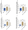
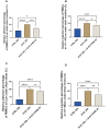
Figure 1
Figure 1
The effects of the severe cytokine cocktail and FX06 on TEM through EC monolayer. PBMC adhesion (0 μm) under static conditions (A) and under shear stress conditions (C), PBMCs migration (-10--100 μm) under static conditions (B) and (-10--50 μm) under shear stress conditions (D) from three independent experiments were measured with the Revvity Harmony software. FX06 reduced the increased TEM caused by the severe cytokine cocktail in ECS cultured under both static and shear stress conditions. One-way ANOVA, ns: non-significant, *P<0.05, **P<0.01, ***P<0.001, ****P<0.0001.
Figure 2
Figure 2
The protective effects of FX06 in ECs treated with the severe cytokine cocktail and S1. PBMC adhesion (0 μm) and migration (-10--100 μm) under static conditions (A, B), PBMC adhesion (0 μm) and migration (-10--50 μm) under shear stress conditions (C, D), from three independent experiments, were measured on Revvity Harmony software. The severe cytokine cocktail together with S1 significantly increased the TEM through ECs monolayer, and FX06 significantly reduced the TEM. One-way ANOVA, ns: non-significant, *P<0.05, **P<0.01, ***P<0.001, ****P<0.0001.
Figure 3
Figure 3
The effects of the severe cytokine cocktail in the absence or presence of FX06 on the viability of ECs. Statistical analysis was conducted on the percentage of alive and early apoptotic cells (A), and the percentage of late apoptotic and necrotic cells (B) from three independent flow cytometric experiments. The severe cytokine cocktail significantly decreased the percentages of alive and early apoptotic cells and increased the percentages of late apoptotic and necrotic cells. FX06 did not reverse the changes caused by the severe cytokine cocktail. One-way ANOVA, ns: not significant, *P<0.05, **P<0.01, ***P<0.001, ****P<0.0001.
Figure 4
Figure 4
The surface expression of CD138 in ECs, treated for 24 h with medium, the severe cytokine cocktail in the absence or presence of 2 h-treatment of FX06. Images of CD138 (A) showed the surface marker in ECS cultured under static and shear stress conditions. Images were taken on Evident Olympus FV1000 with 40x objective. The surface expression of CD138 in ECs cultured under both static and shear stress conditions (B, C) was measured via immunofluorescence. Under both static and shear stress conditions, the severe cytokine cocktail caused more surface expression of CD138, and FX06 reversed the surface expression level of CD138 back to control levels. (D) shows the level of shed CD138. The severe cytokine cocktail induced more shed CD138, whereas FX06 treatment caused even more. One-way ANOVA, ns, not significant, *P<0.05, **P<0.01, ***P<0.001, ****P<0.0001.
Figure 5
Figure 5
The morphological changes of ECs. The cell area (A), cell length (B), and cell width (C) were measured with the Revvity Harmony software. One-way ANOVA, ns: not significant, *P<0.05, ****P<0.0001. (D) ECs were treated with medium, the severe cytokine cocktail, and the severe cytokine cocktail in the presence of 2h-treatment of FX06. ECs were incubated with anti-F-actin antibodies, Alexa Fluor 488 Phalloidin (green). DAPI (blue) was utilised to stain nuclei. Images were taken with a Revvity Phenix Opera Confocal Microscope using 40x water immersion objective. Scale bar: 50 μm.
Figure 6
Figure 6
The effects of the severe cytokine cocktail and the severe cytokine cocktail with the presence of FX06 on angiogenesis activities in ECs. Images from (A) were taken from Zeiss Primovert microscope with a 4x objective. Scale bar: 500 μm. The total number of nodes and junctions (B), the number of meshes (C), and the total number of segments and branches (D), from five independent experiments were measured by angiogenesis analyzer on ImageJ. One-way ANOVA, ns: not significant, *P<0.05, **P<0.01.
Figure 7
Figure 7
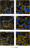
The effects of the severe cytokine cocktail and the severe cytokine cocktail with the presence of FX06 on VE-cadherin distribution in ECs. ECs were incubated with anti-VE-cadherin antibodies and stained with Alexa Fluor 546 (yellow). DAPI (blue) was utilised to stain nuclei. ECs were treated with medium, the severe cytokine cocktail, and the severe cytokine cocktail in the presence of 2 h-treatment of FX06. Images (A-C) were taken with a Revvity Phenix Opera Confocal Microscope using 40x water immersion objective. Scale bar: 50 μm. White boxes highlight the zoomed areas (a-c). Zoomed scale bar: 18.75 μm.
Figure 8
Figure 8
The cytotoxicity of CD8+ T cells on ECs. The percentages of ECs specific cell lysis caused by CD8+ T cells were measured by flow cytometry analysis. (A-C) The flow cytometry analysis gating strategy. The graphs were from one sample. First singlets were gated (A), then DIO+ cells were gated in singlets (B). At the end PI+ population that represented CD8+ T cells-induced EC lysis was gated in DIO+ cells (C). Same gatings were applied to all samples with different treatment. (D) A549 CTRL served as a negative control. Severe cytokine cocktail treatment increased the cell lysis which was downregulated by FX06 treatment. One-way ANOVA, ns: not significant, **P<0.01.
Figure 9
Figure 9
(A) The experimental design of proteomics and phosphoproteomics analysis. The illustration was created by Biorender. (B) Box plots with the overall number of upregulated and downregulated proteins (top) and phosphosites (bottom) for six cell group comparisons including FX06 treatments (5, 20, 60, 120, 360 min) versus Severe and Severe versus Control. (C) Volcano plots of differentially expressed proteins and red circles depict significantly differentially expressed proteins. FX06 induces differentially expressed proteins over the time. (D) Bubble plot with the results of Gene Set Enrichment Analysis using the KEGG database. The severe cytokine cocktail with the addition of 2h-FX06 shows the protective effects on EC barrier integrity.
Figure 10
Figure 10
(A) MFuzz clustering. Line charts with changes in protein phosphorylation for each phosphorylation site in its respective cluster at each time point of the experiment. Lines with yellow, green, and blue colours indicate proteins with low membership values, while the lines with red and purple colours correspond to proteins with high membership values. The black line highlights the centre of the cluster at each time point of the experiment. The addition of FX06 to the severe cytokine cocktail has shown to normalize the changesinduced by the severe cytokine cocktail. (B) Heatmaps with the results of Over-representation analysis using the Reactome databases. The reversed effects of FX06 in Rho GTPase are observed. (C) Heatmap with Kinase Enrichment Scores from KSEA based on whole phosphoproteomics data. Kinases with Enrichment Scores with FDR values less than 0.05 are marked with the "*" symbol. Only kinases with p-value < 0.05 are selected. CSNK2A1 is downregulated by FX06 at all time points.
Figure 11
Figure 11
(A) DIABLO sample plot based on sample scores on first (Variable 1) and second (Variable 2) components from proteomics and phosphoproteomics data. (B) Overrepresentation analysis of top 100 proteomics and phosphoproteomics loadings of component 2 based on Reactome database. Only statistically significant pathways are selected (p-value < 0.05). The length of bars represents -log10 (p-value).
Figure 12
Figure 12
The effects of the severe cytokine cocktail alone, and the severe cytokine cocktail in the presence of FX06 on the total expression of RhoA and phospho-ROCK1 in HULEC-5a (C, D) Protein lysates were collected for Western blots to measure RhoA and phospho-ROCK1 with an anti-RhoA antibody and anti-Phospho-ROCK1 (Thr455, Ser456). Anti-vinculin and anti-GAPDH as a housekeeping marker were used to normalise data. A representative blot of three independent experiments is shown above. (A, B) Western bands were quantified via ImageJ. Quantification data from three and six independent experiments regarding RhoA and phospho-ROCK1 expression is shown in the graph. 2 h-treatment of FX06 decreased the RhoA expression and phospho-ROCK1 that was enhanced by the severe cytokine cocktail. One-way ANOVA, ns: not significant, *P<0.05, ***P<0.001, ****P<0.0001.

References

    1. Appelman B, Charlton BT, Goulding RP, Kerkhoff TJ, Breedveld EA, Noort W, et al. Muscle abnormalities worsen after post-exertional malaise in long COVID. Nat Commun. (2024) 15:17. doi: 10.1038/s41467-023-44432-3 - DOI - PMC - PubMed
    1. Greene C, Connolly R, Brennan D, Laffan A, O’Keeffe E, Zaporojan L, et al. Blood–brain barrier disruption and sustained systemic inflammation in individuals with long COVID-associated cognitive impairment. Nat Neurosci. (2024) 27(3):421–32. doi: 10.1038/s41593-024-01644-0 - DOI - PMC - PubMed
    1. Cimino G, Vizzardi E, Calvi E, Pancaldi E, Pascariello G, Bernardi N, et al. Endothelial dysfunction in COVID-19 patients assessed with Endo-PAT2000. Monaldi Arch Chest Dis. (2022) 92(4). doi: 10.4081/monaldi.2022.2213 - DOI - PubMed
    1. Bourgonje AR, Abdulle AE, Timens W, Hillebrands J, Navis GJ, Gordijn SJ, et al. Angiotensin-converting enzyme 2 ( ACE2 ), SARS-CoV -2 and the pathophysiology of coronavirus disease 2019 ( COVID -19). J Pathol. (2020) 251:228–48. doi: 10.1002/path.v251.3 - DOI - PMC - PubMed
    1. Cabaro S, D’Esposito V, Di Matola T, Sale S, Cennamo M, Terracciano D, et al. Cytokine signature and COVID-19 prediction models in the two waves of pandemics. Sci Rep. (2021) 11:20793. doi: 10.1038/s41598-021-00190-0 - DOI - PMC - PubMed

MeSH terms