. 2025 Oct;37(39):e2504519.
doi: 10.1002/adma.202504519. Epub 2025 Jul 4.

Flippase-Mediated Hybrid Vesicle Division

Affiliations

Flippase-Mediated Hybrid Vesicle Division

Paula De Dios Andres et al. Adv Mater. 2025 Oct.

Abstract

The assembly of synthetic systems with the ability for protein-mediated division remains a challenge in bottom-up synthetic biology. Here, the reconstitution of an active Drs2p-Cdc50p lipid flippase in polymer lipid hybrid vesicles (HVs) made from phospholipids and 1 or 2.5 mol% amphiphilic block copolymers, with poly(carboxyethyl acrylate) or poly(6-O-methacryloyl-d-galactopyranose) as the hydrophilic extension and either cholesteryl methacrylate or butyl methacrylate or combinations thereof as the hydrophobic blocks is demonstrated. The reconstitution of Drs2p-Cdc50p in HVs flip 2-dioleoyl-sn-glycero-3-phospho-l-serine (DOPS) lipids from the inner to the outer leaflet, leading to transmembrane asymmetry. Importantly, the chemical nature of the hydrophobic block in the amphiphilic block copolymers used to assemble the HVs is crucial to support changes in the spontaneous curvature of the bilayers due to translocation of DOPS lipids that results in HV constriction and division. Taken together, this effort is a step forward in imitating cell division in synthetic assemblies toward potentially bottom-up assembled self-replicating units.

Keywords: hybrid vesicle; lipid flippase; membrane constriction; transmembrane asymmetry; vesicle division.


Conflict of interest statement

The authors declare no conflict of interest.

Figures

Scheme 1
Scheme 1
a) Cartoon showing HVs with reconstituted Flippase (HVXFlip) that undergoes constriction and division when exposed to ATP due to the translocation (flipping) of the DOPS lipids. b) i) Schematic illustration of three different amphiphilic block copolymers (BCPs), with different hydrophobic blocks and either poly(carboxyethyl acrylate) or poly(6‐O‐methacryloyl‐d‐galactopyranose) hydrophilic extension. ii) The hydrophobic block in poly(cholesteryl methacrylate)‐block‐poly(2‐carboxyethyl acrylate) (BCP1) is poly(cholesteryl methacrylate), poly(cholesteryl methacrylate‐co‐butyl methacrylate) in poly(cholesteryl methacrylate‐co‐butyl methacrylate)‐block‐poly(2‐carboxyethyl acrylate) (BCP2) as well as poly(cholesteryl methacrylate‐co‐butyl methacrylate)‐block‐poly(6‐O‐methacryloyl‐D‐galactopyranose) (BCP4), and poly(butyl methacrylate) in poly(butyl methacrylate)‐block‐poly(2‐carboxyethyl acrylate) (BCP3). iii) BCPX can be employed to assemble small or giant hybrid vesicles (sHVX or GHVX) together with phospholipids DOPS and 1,2‐dioleoyl‐sn‐glycero‐3‐phosphocholine (DOPC) as well as1,2‐dioleoyl‐sn‐glycero‐3‐phospho‐(1′‐myo‐inositol‐4′‐phosphate) (PI4P) for reconstitution with Flippase. iv) Illustration of the Flippase activity in the hybrid membrane using DOPS as substrate.
Figure 1
Figure 1
Flippase reconstitution in sHVXs. a) Illustration showing the Flippase reconstitution process in sHVXs. Representative negative‐stain TEM (top) and cryoEM (bottom) images of b) sHV1, c) sHV2, and d) sHV3 only exposed to CHAPS, sHVXC (left) or exposed to CHAPS to reconstituted Flippase, sHVXFlip (right). Insets in the upper right corners show a 2.5× zoom (scale bar: 500 nm (top)/50 nm (bottom)).
Figure 2
Figure 2
a) i) ATP consumption of sHVXFlip in comparison to sHVXC. sHVXFlip without ATP addition did not give any signal. ATP consumption of sHV2Flip ii) without PI4P, and iii) without DOPS lipids. The data were normalized to the value of fluorescence intensity of 1 mM ATP (final concentration) and presented as normalized fluorescence intensity (nFI). b) Repeated ATP consumption of sHVXFlip over 3 h. The arrows indicate the ATP addition (n = 3; the data are expressed as mean ± standard deviation (SD)).
Figure 3
Figure 3
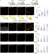
a) Schematic illustration showing the ATP‐dependent DOPS lipid redistribution to the outer leaflet in sHVXFlip followed by the immobilization of AnnexinF. Representative super‐resolution images of b) sHV1, c) sHV2, and d) sHV3, (i) sHVXC − ATP, ii) sHVXC + ATP, iii) sHVXFlip − ATP, and iv) sHVXFlip + ATP (orange: AnnexinF; scale bar: 5 µm)), to and v) the corresponding pixel quantification of AnnexinF expressed as AnnexinF area in %, indicating the area above the threshold (n = 2; the data are expressed as mean ± SD * p < 0.1; ** p < 0.05, one‐way analysis of variance (ANOVA) with Šídák's multiple‐comparisons test, the * above a plot indicates a significant difference from all other groups).
Figure 4
Figure 4
Flippase reconstitution in GHVs. i) Representative overview CLSM images of a) GHV2Flip, b) GHV3Flip, c) GHV4Flip, and d) GHV4Flip2.5 (green: Oregon Green‐labeled BCPX, red: Rho‐PE, blue: FlippaseF) (scale bar: 20 µm). CLSM images of a ii) representative GHVXFlip vesicle and a iii) representative lipid only based giant vesicle from the same population split in images with the signal originating from Rho‐PE (top), OGBCPX (middle), and FlippaseF (bottom). iv) ATP consumption of GHVXFlip and pristine GHVXC in comparison to GHVXFlip in the absence of ATP (n = 3; the data are expressed as mean ± SD).
Figure 5
Figure 5
a) Schematic illustration of the assessment of the transmembrane distribution of NBD‐PS in GHV2Flip when preincubated with ATP before exposure to dithionite. b) Representative CLSM images of i) GHV2Flip + ATP, ii) GHV2Flip − ATP, iii) GHV2C + ATP, and iv) GHV2C ‐ ATP. c) Whisker plot of the fluorescence intensity of the membrane of GHV2c or GHV2Flip before (black) and after (gray) dithionite exposure depending on the ATP preincubation (green: NBD‐PS; scale bar: 10 µm; n = 2, N = 400).
Figure 6
Figure 6
Transmembrane distribution of DOPS lipids in GHV2Flip. a) Schematic illustration showing the ATP‐dependent DOPS lipid redistribution to the outer leaflet in GHV2Flip followed by the binding of AnnexinF. b) i) Representative CLSM images and ii) the corresponding whisker plot of the vesicles fluorescence intensities of GHV2C ‐ ATP, GHV2C + ATP, GHV2Flip − ATP, and GHV2Flip + ATP after incubation with AnnexinF. iii) Pie charts showing the ATP‐dependent distribution of constricted (“snowman‐” like) and spherical GHV2C and GHV2Flip observed in CLSM images. c) i) Representative CLSM and ii) the corresponding whisker plot of the vesicles fluorescence intensities of GHV3C ‐ ATP, GHV3C + ATP, GHV3Flip − ATP, and gHV3Flip + ATP. d) i) Representative CLSM images and ii) the corresponding whisker plot of the vesicles fluorescence intensities of GHV4C ‐ATP, GHV4C + ATP, GHV4Flip − ATP, and GHV4Flip + ATP. e) i) Representative CLSM images and ii) the corresponding whisker plot of the vesicles fluorescence intensities of GHV4C2.5 ‐ ATP, GHV4C2.5 + ATP, GHV4Flip2.5 − ATP, and GHV4Flip2.5 + ATP (orange: AnnexinF; scale bar: 10 µm) (n = 2, N = 400, * p < 0.05, one‐way ANOVA with Šídák's multiple‐comparisons test, the * above a plot indicates a significant difference from all other groups).
Figure 7
Figure 7
a) Schematic of GHV2Flip exposed to ATP leading to constriction (top) or constriction followed by division (bottom). b) Representative CLSM image series of constricted GHV2Flip after 20 to 60 min exposure to ATP, and of (c) dividing gHV2Flip 30 to 60 min after ATP addition. d) Illustration of GHV4Flip in the presence of ATP. e) Representative CLSM image series of constricted GHV4Flip after 10 to 15 min exposure to ATP (red: Rho‐PE, scale bars: 10 µm).

References

    1. a) Schwille P., Spatz J., Landfester K., Bodenschatz E., Herminghaus S., Sourjik V., Erb T. J., Bastiaens P., Lipowsky R., Hyman A., Dabrock P., Baret J.‐C., Vidakovic‐Koch T., Bieling P., Dimova R., Mutschler H., Robinson T., Tang T.‐Y. D., Wegner S., Sundmacher K., Angew. Chem., Int. Ed. 2018, 57, 13382; - PubMed
    2. b) Mulla Y., Aufderhorst‐Roberts A., Koenderink G. H., Phys. Biol. 2018, 15, 041001; - PubMed
    3. c) Stano P., Life 2019, 9, 3.
    1. Yewdall N. A., Mason A. F., van Hest J. C. M., Interface Focus 2018, 8, 20180023. - PMC - PubMed
    1. Chang M.‐Y., Ariyama H., Huck W. T. S., Deng N.‐N., Chem. Soc. Rev. 2023, 52, 3307. - PubMed
    1. Chemin M., Brun P.‐M., Lecommandoux S., Sandre O., Meins J.‐F. L., Soft Matter 2012, 8, 2867.
    1. Qutbuddin Y., Guinart A., Gavrilović S., Al Nahas K., Feringa B. L., Schwille P., Adv. Mater. 2024, 36, 2311176. - PubMed

MeSH terms

LinkOut - more resources