. 2025 Dec;19(1):2517851.
doi: 10.1080/19336950.2025.2517851. Epub 2025 Jul 4.

A rare HCN4 variant combined with sick sinus syndrome, left ventricular noncompaction, and complex congenital heart disease

Affiliations

A rare HCN4 variant combined with sick sinus syndrome, left ventricular noncompaction, and complex congenital heart disease

Fengxiao Zhang et al. Channels (Austin). 2025 Dec.

Abstract

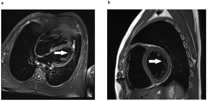
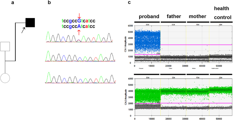
The hyperpolarization-activated cyclic nucleotide-gated potassium channel 4 (HCN4) gene has been reported to regulate the spontaneous depolarization of sinoatrial node cells. A novel HCN4 mutation (c.2036 G>A) may lead to sick sinus syndrome. The green fluorescent protein (GFP) and either the wild-type (WT) or C679Y mutant (mut) were co-transfected into HEK293 cells to investigate the impact of the mutation on HCN4 channel function. The whole-cell patch-clamp approach was utilized to record HCN4 currents. According to electrophysiological recording, the current amplitude and density generated by mut-C679Y HCN4 channels were much lower than those generated by WT channels. HCN4 channel current activation was not significantly affected by the C679Y mutation. Because of the little current, analyzing the mut channel deactivation kinetic was challenging. Thus, we have identified a novel HCN4 gene mutation that is connected to bradycardia, left ventricular noncompaction, and diverse valve-related heart conditions.

Keywords: HCN4; Sick sinus syndrome; left ventricular noncompaction.


Conflict of interest statement

No potential conflict of interest was reported by the author(s).

Figures

Electrocardiograph of the proband. This ECG report indicates bradycardia, with no other abnormalities.
Figure 1.
Electrocardiograph of the proband. The ECG showed sinus bradycardia with a heart rate of 47 beats.
These are two echocardiographic (ultrasound of the heart) images, labeled as a and b a. It shows a cross-sectional view of the heart. The numbered structures (1, 2, 3, 4) likely correspond four cusps of the aortic valve. The positions of four different valve blades are numerically identified. b. Displays another cross - sectional view of the heart. The positions of three different valve blades are numerically identified.
Figure 2.
Echocardiographic image of the patient with combined malformation of aortic valve and pulmonary valve. (a). Short-axis transesophageal echocardiogram (TEE) image shows four cusps of the aortic valve. (b). Short-axis TEE image shows more than three cusps of the pulmonary valve.
Cardiac magnetic resonance of the patient with LVNC. Fat-suppressed T2-weighted imaging shows a non-compacted myocardium. Incomplete densification occurred in the free wall of the left ventricle and the myocardium at the apex. The thickness ratio of dense myocardium to non-dense myocardium is less than 2:1. Arrowhead marks the site of non-compacted myocardium.
Figure 3.
Cardiac magnetic resonance of the patient with LVNC. Fat-suppressed T2-weighted imaging shows a non-compacted myocardium.
Figure 4.
Figure 4.
(a). Family tree of the proband. Squares represent male family members and circles represent female family members. The proband is represented by a black square. (b). Sanger sequencing validation of the HCN4 mutation in blood samples. The C679Y mutation is denoted by the red arrowheads. Samples are numbered as follows: 1, proband; 2, father; and 3, mother. (c). Fluorescence intensity of the droplets following amplification of the HCN4 mut region using the ddPCR expert design assays (Bio-Rad laboratories). The four ddPcrs are divided by vertical dotted yellow lines for the proband, mother, father, and health control. The pink line is the threshold, above which are positive droplets (blue and green), and below which are negative droplets (gray) without any target DNA. There is no target DNA for the mutant locus c.2036 G>A in the mother and father.
Figure 5.
Figure 5.
I-V relationship and activation and deactivation properties of C679Y. (a). Voltage protocol and representative current traces were recorded from HEK293 cells expressing wild-type (WT) and C679Y HCN4 channels. (b). Resulting current density amplitude is plotted against voltage. (c). Current densities recorded at −120 mV membrane potential. (d). Reversal potential mean values for WT and C679Y channels. The reversal potential was computed utilizing a third-order polynomial function. (e). Activation pulse protocol (above) of the activation curve and the representative whole-cell current traces of HEK293 cells expressing HCN4. (f). Deactivation pulse protocol (above) and the representative whole-cell current traces of HEK293 cells expressing WT and C679Y HCN4 channels. T two-tailed unpaired Student’s t-test was used for the statistical difference analysis.
a and b show the predicted structures of wild - type and C679Y mutant HCN4 proteins, respectively. Amino acids 521 - 713 are colored green, and the CNBD (cyclic nucleotide - binding domain) region spanning 602 - 720 is in brown. The additional small molecule represents cAMP. In a, the wild - type protein has Cysteine at position 679 (C679), while in b, the mutant protein has Tyrosine at position 679 (Y679) due to the C679Y mutation.
Figure 6.
Crystal structure of the WT-HCN4 (a) and mut-HCN4 (b) proteins predicted by software Alphafold 2. The amino acid residues from position 521 to 713 of the HCN4 protein are marked in green. The region (the 602nd to the 720th amino acid residues) encompasses the cyclic nucleotide-binding domain (CNBD), which is marked in brown. The 679th amino acid residue is highlighted in red, which is cysteine in wt-HCN4 (a) and tyrosine in mut-HCN4 (b). The small molecule represents cAMP.

References

    1. DiFrancesco D. The role of the funny current in pacemaker activity. Circ Res. 2010. Feb 19;106(3):434–11. doi: 10.1161/CIRCRESAHA.109.208041 PubMed PMID: 20167941. - DOI - PubMed
    1. Zagotta WN, Olivier NB, Black KD, et al. Structural basis for modulation and agonist specificity of HCN pacemaker channels. Nature. 2003. Sep 11;425(6954):200–205. doi: 10.1038/nature01922 PubMed PMID: 12968185. - DOI - PubMed
    1. Mengesha HG, Tafesse TB, Bule MH. I(f) channel as an emerging therapeutic target for cardiovascular diseases: a review of current evidence and controversies. Front Pharmacol. 2017;8:874. doi: 10.3389/fphar.2017.00874 PubMed PMID: 29225577; PubMed Central PMCID: PMCPMC5705549. - DOI - PMC - PubMed
    1. Verkerk AO, Wilders R. Pacemaker activity of the human sinoatrial node: effects of HCN4 mutations on the hyperpolarization-activated current. EP Europace. 2014. Mar;16(3):384–395. doi: 10.1093/europace/eut348 PubMed PMID: 24569893. - DOI - PubMed
    1. DiFrancesco D. HCN4, sinus bradycardia and atrial fibrillation. Arrhythm Electrophysiol Rev. 2015. May;4(1):9–13. doi: 10.15420/aer.2015.4.1.9 PubMed PMID: 26835093; PubMed Central PMCID: PMCPMC4711571. - DOI - PMC - PubMed

MeSH terms

Substances