. 2025 Jun 20;53(12):gkaf574.
doi: 10.1093/nar/gkaf574.

TRIP12's role in the governance of DNA polymerase β involvement in DNA damage response and repair

Affiliations

TRIP12's role in the governance of DNA polymerase β involvement in DNA damage response and repair

Burcu Inanc et al. Nucleic Acids Res. .

Abstract

The multitude of DNA lesion types, and the nuclear dynamic context in which they occur, presents a challenge for genome integrity maintenance as this requires the engagement of different DNA repair pathways. Specific "repair controllers" that facilitate DNA repair pathway crosstalk between double-strand break (DSB) repair and base excision repair (BER) and that regulate BER protein engagement at lesion sites have yet to be identified. Here, we find that DNA polymerase β (Polβ), crucial for BER, is ubiquitylated in a BER complex-dependent manner by TRIP12, an E3 ligase that partners with UBR5 to restrain DSB repair signaling. Furthermore, we find that TRIP12, but not UBR5, controls cellular levels and chromatin loading of Polβ. Required for Polβ foci formation, TRIP12 influences Polβ involvement after radiation-induced DNA damage, a process regulated by TRIP12-mediated ubiquitylation of Polβ. Notably, excessive TRIP12-mediated engagement of Polβ affects DSB formation and radiation sensitivity, underscoring its role in promoting precedence for BER over DSB repair. The herein discovered function of TRIP12, in the governance of Polβ-directed BER, supports a role for TRIP12 in assuring BER lesion removal at complex DSB sites to optimize DSB repair at the nexus of DNA repair pathways.


Conflict of interest statement

None declared.

Figures

Graphical Abstract
Graphical Abstract
Figure 1.
Figure 1.
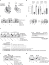
TRIP12 as a novel Polβ partner. (A) BER complex compositions and differential partner identification strategy. The graphic depicts the immunoprecipitation scheme used to discover novel interacting BER protein partners. (B) TRIP12 is a novel Polβ binding partner with an increased affinity to the XRCC1-free complex (Complex B). Quantification of selected proteins bound to Complex A or Complex B, as indicated, by label-free dMS. The relative abundances of prototypic peptides with unique amino acid sequences AIGSTSKPQESPK, SEAHTADGISIR, VNNGNTAPEDSSPAK, and LSTQSNSNNIEPAR were used as surrogate measures of XRCC1, LIG3, PARP2, and TRIP12, respectively. Peptide abundance levels were normalized to Polβ in each immunoprecipitated sample and are shown as dots on bar plots with mean and SD. Significant differences between complexes A and B are marked by asterisks (***P< .001, ****P< .0001; ANOVA). (C) BER complex-dependent interaction of TRIP12. Interaction of TRIP12 with Flag-Polβ(WT) or Flag-Polβ(TM) was revealed by IP from whole cell lysates (WCLs) and IB as indicated. (D) Interaction of endogenous Polβ with TRIP12. IP/IB of endogenous proteins in the indicated cell lines are shown. (E) Domain interaction mapping. Flag-Polβ binding to myc-TRIP12 domains as depicted in the scheme shown to the right and determined by IP/IB: myc-TRIP12 (full length TRIP12, with an N-terminal myc-tag), myc-TRIP12-SB (TRIP12 SB domain, amino acids 1–1650, with an N-terminal myc-tag), and myc-TRIP12-HECT (TRIP12-HECT domain, amino acids 1651–2040, with an N-terminal myc-tag). (F) Domain interaction mapping of myc-TRIP12 to the C-terminal domain of Polβ wild-type and mutants by IP/IB: Flag-Polβ(WT)-C (amino acids 91–335), Flag-Polβ(TM)-C, XRCC1 binding mutant (amino acids 91–335), and Flag-Polβ(TM/DM)-C, ubiquitylation and XRCC1 binding mutant (amino acids 91–335). (G) Domain interaction mapping of myc-TRIP12 to the N-terminal domain of Polβ wild-type and mutants by IP/IB: Flag-Polβ(WT)-N (amino acids 1–90).
Figure 2.
Figure 2.
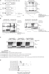
TRIP12’s role in Polβ ubiquitylation and degradation. (A) Ex vivo Polβ ubiquitylation by TRIP12. Polβ ubiquitylation by TRIP12 as identified by immunoblot (right) after the “on-bead” Ubi- assay as outlined in the scheme (left) using recombinant and purified His-ubiquitin, E1/E2, and Polβ together with myc-HECT, myc-HECT(C2007A), myc-TRIP12-SB, or myc-TRIP12 expressed in and isolated from LN428 cells. Boxes indicate the fractions analyzed (FR1–FR4). Immunoblot of FR4 (right) indicates that Polβ is ubiquitylated by full-length myc-TRIP12 or the HECT domain but not the substrate-binding domain or the HECT domain with the active site mutation (C2007A). (B) TRIP12-HECT-dependent ubiquitylation. Immunoblots as in panel (A), using FR3 and FR4 from the Ubi assay as indicated in panel (A) with myc-HECT (wild-type or C2007A mutant) after different incubation times. (C) TRIP12-dependent ubiquitylation of transgenic Polβ in cells. Ubiquitylation of Flag-tagged wild-type Polβ [Flag-Polβ(WT)], XRCC1-binding mutant Polβ(L301R/V303R/V306R) [Flag-Polβ(TM)], and of the ubiquitylation and XRCC1-binding mutant Polβ(L301R/V303R/V306R/K206A/K244A) [Flag-Polβ(TM/DM)] was determined by IP/IB following transfection of HA-ubiquitin and using two different shRNA (sh1 and sh5) to TRIP12. Flag-IP HA antibody blotted lanes show ubiquitylation of pulled down proteins of which the majority are Polβ and Polβ complex-associated proteins. These are compared to IP input (WCL extracts) and probed for expression of PCNA (input loading control), Polβ, and HA (Ub). (D) TRIP12 affects cellular Polβ levels. Shown are changes (fold increase) in Polβ isoform levels, as listed [Flag-Polβ(WT), Flag-Polβ(TM), or Flag-Polβ(DM)], in TRIP12-depleted cells (by TRIP12-sh1 or sh5 knockdown) when compared to their respective control-shRNA (SCR) expressing LN428 cells. Bar graph depicts average control-shRNA (SCR) normalized Polβ isoform levels as determined by multiple immunoblots (representative IB in Supplementary Fig. S2D), each indicated by a dot, with SD and n = 3–4. Asterisks indicate statistically significant differences (**P< .01, ****P< .0001; ANOVA) to the corresponding control-shRNA results.
Figure 3.
Figure 3.
TRIP12-mediated Polβ chromatin retention and BER function (A) Prominent nuclear localization of TRIP12. Confocal microscopy images showing nuclear (blue) localization of TRIP12 (green). (B) TRIP12-mediated Polβ chromatin retention. Immunoblots show Polβ, XRCC1, tubulin (cytosol fraction loading control), and SSRP1 (chromatin fraction loading control) levels in WCLs, the cytosolic or chromatin fraction in two different TRIP12-KD (knockdown) cell lines (TRIP12-sh1 and -sh5), scrambled controls (C), and the parental LN428 cell line (–) as indicated. (C) Laser damage induced focal recruitment of Polβ, independent of TRIP12. Quantification of laser (405 nm)-induced local recruitment and retention of copGFP fused Polβ (copGFP-Polβ) in TRIP12 knockdown (TRIP12-sh1 and -sh5) and control LN428 cell lines. Data are from n = 2 independent experiments with each n = 10 cells. (D) Effective oxidative damage repair in TRIP12-depleted cells. Oxidative damage and repair as determined by alkaline CometChip analyses (tail DNA in %) following a 30-min exposure to 250 μM H2O2 (damaged) and at 60 min post-exposure (repair) in TRIP12-KD (TRIP12-sh1 and -sh5), scrambled (SCR), or parental (WT) control cell lines. (E) Cellular response to oxidative damage is not affected by the loss of TRIP12. H2O2 sensitivity of TRIP12-depleted cell lines (TRIP12-sh1 and -sh5) is not significantly different from scrambled controls (SCR) as determined by clonogenic survival and curve fit comparisons or H2O2 IC50 determinations (ANOVA). Shown are the mean surviving fractions of three independent experiments ± SD. (F) Depletion of TRIP12 does not affect cellular survival. No significant changes (ANOVA) were observed in the clonogenic survival of TRIP12-depleted cell lines (TRIP12-sh1 and -sh5), scrambled controls (SCR), and parental LN428 cells as determined by colony formation assays. The mean of three to four independent experiments, each indicated by dots with ± SD, is shown. (G) Depletion of TRIP12 does not affect cell growth. TRIP12-depleted cell lines (TRIP12-sh1 and -sh5), LN428 parental cells (WT), and scrambled control (SCR) growth as determined by the MTT assay (mean and SD of n = 4).
Figure 4.
Figure 4.
TRIP12 promotes radiation-induced Polβ foci formation. (A) Polβ foci formation by radiation. Representative images of copGFP-fused Polβ (copGFP-Polβ) foci in LN428 cells at different time points after radiation (10 Gy). (B) Characterization of radiation-induced Polβ foci over time. Dot plots show the distribution of copGFP-Polβ foci in untreated cells (Unt) and in cells at 5 min, 1 h, or 5 h after 10 Gy (n = 3 independent experiments with >50 cells each). Mean foci/cell values per experiment and corresponding statistical evaluations are shown in Supplementary Fig. S4A and B. ****P< .001 in the Kruskal–Wallis test. (C) Polβ foci formation is radiation dose dependent. Average copGFP-Polβ foci counts per cell are shown of all foci (left Y axis and blue values) and of large foci (>8 px, right Y axis with purple values) at 1 h after radiation. Data show mean and SD of pooled foci counts of two to three independent experiments and ****P< .001 indicate multiple comparisons adjusted to the respective unirradiated controls (ANOVA). (D) Polβ foci colocalize with XRCC1. Changes in mean Polβ(WT) foci/cell and Polβ(WT)/XRCC1 colocalized foci of n = 3 independent experiments are shown with *P< .05, **P< .01, ***P< .001, and ****P< .0001 as determined by ANOVA. (E) Reduced Polβ foci and XRCC1 colocalization of Polβ isoforms devoid of XRCC1 binding [copGFP-Polβ(TM) or copGFP-Polβ(TM/DM)]. Violin plots show copGFP-Polβ isoform foci [copGFP-Polβ(WT), copGFP-Polβ(TM), and copGFP-Polβ(TM/DM)], XRCC1 foci in copGFP-Polβ(WT), copGFP-Polβ(TM), and copGFP-Polβ(TM/DM) expressing cells, and copGFP-Polβ isoform/XRCC1 colocalized foci distributions. Kruskal–Wallis test results are shown and interexperimental variations are shown in Supplementary Fig. S4C and D; representative images are shown in Supplementary Fig. S4E. (F) Majority of Polβ foci do not colocalize with γH2AX foci. Numbers of copGFP-Polβ(WT), γH2AX, and Polβ(WT)/gH2AX colocalized foci of individual cells (left) or mean foci/cell values per experiment (right) unirradiated and following 5 h after 10 Gy irradiation are shown; *P< .05, **P< .01, ***P< .001, and ****P< .0001 as determined by Kruskal–Wallis (left) or ANOVA comparing the average of the means from three independent experiments (right). (G) Polβ foci do not colocalize with 53BP1 foci. Numbers of copGFP-Polβ(WT), 53BP1, and Polβ(WT)/53BP1 colocalized foci of individual cells (left) or mean foci/cell values per experiment (right) unirradiated and following 10 Gy irradiation and 5-h repair are shown; *P< .05, **P< .01, ***P< .001, and ****P< .0001 as determined by Kruskal–Wallis (left) or ANOVA comparing the average of the means from three independent experiments (right). (H) Representative images illustrating the quantified colocalization pattern of Polβ and XRCC1 foci or lack thereof with 53BP1. (I) TRIP12-mediated Polβ foci formation. Representative images of TRIP12-controlled Polβ recruitment after radiation (10 Gy) in scrambled (SCR) control cells and TRIP12-depleted (TRIP12-sh1 and -sh5) cells. (J) Late and large Polβ foci and Polβ foci rich cells are particularly affected by TRIP12 depletion. Graph shows quantification of radiation-induced Polβ [copGFP-Polβ)] foci in scrambled (SCR) control and TRIP12-sh1 and -sh5 cells as indicated. Data show the number of large (>8 px) Polβ foci per cell from n = 3 independent experiments with >50 analyzed cells each; bars indicate means with SD. Asterisks mark multiple comparison adjusted P-values (****P< .0001) in the Kruskal–Wallis test comparing the different cell line results at each time point to each other. (K) Robust abrogation of radiation-induced Polβ foci by TRIP12 depletion. Quantification of radiation-induced Polβ [copGFP-Polβ)] foci in scrambled (SCR) control and TRIP12-sh1 and -sh5 cells as in panel (J). Graph demonstrates the interexperimental variation not visible in panel (J) and shows the average and SD of the means from n = 3 independent experiments over time. Control SCR data used as reference; asterisks indicate significantly different mean foci numbers with *P< .05, ***P< .00, and ****P< .0001 (ANOVA). ANOVA reports a significant interaction with P< 0.05. Radiation-induced foci are significantly different from untreated, only in the SCR for the 1 and 5 h data points with P< .001 and P< .0001, respectively.
Figure 5.
Figure 5.
Repair interference by forced Polβ imbalance (A) Radiation response is unaltered by TRIP12 depletion. TRIP12-depleted cells (TRIP12-sh1 and -sh5) were compared to scrambled (SCR) LN428 control cells. Clonogenic survival after 4 Gy with n = 2–3 independent experiments; errors are SD; and ns = non-significant ANOVA test results. (B) Polβ overexpression results in increased residual DSBs. Forced Polβ shuttling by excess Polβ results in increased residual γH2AX and 53BP1 foci per cell 24 h after radiation (4 Gy). Polβ overexpressing LN428 cells [ov-Polβ(WT)] were compared to empty vector controls (EV-control). Dot plots show foci per cell counts of n = 3 independent experiments with minimal 50 cells each: *P< .05 and ****P< .0001 (ANOVA). (C) Radiosensitization by deregulated Polβ. Cellular survival (clonogenicity as plating efficiency) and survival after radiation drops in Polβ overexpressing cells [ov-Polβ(WT)] to the same extent as in XRCC1-depleted (XRCC1-KD) cells. Bars and values show the mean and SD of the averages from three to seven independent experiments as indicated by the dots. P-values indicated in the radiation response curve graphs assess the likelihood of the data curve fits to be similar. Radiation response parameters (D37%) in the ov-Polβ(WT) and XRCC1-KD differ significantly from the reference (ref) EV-control cell line with P< .01 and P< .01 (ANOVA), respectively.
Figure 6.
Figure 6.
TRIP12 influences radiation response through Polβ ubiquitylation. (A) TRIP12 depletion reduces Polβ overexpression-induced DSB formation after radiation. Numbers of γH2AX foci per cell in copGFP-Polβ overexpressing scrambled (SCR) control cells and TRIP12-depleted (TRIP12-sh1 and -sh5) cells are shown, unirradiated and 5 h following 10 Gy irradiation as indicated; *P< .05, **P< .01, ***P< .001, and ****P< .0001 as determined by Kruskal–Wallis. (B) TRIP12 depletion rescues cells from Polβ overexpression-mediated radiosensitization. Reduced survival after radiation by the overexpression of wild-type Polβ [ov-Polβ(WT)] (Fig. 5C) in empty vector (EV-control) or SCR (scrambled shRNA) controls is rescued by TRIP12 depletion (TRIP12-sh1 and -sh5). The graph shows the average clonogenic survival after 4 Gy and the SD in n = 3–7 independent experiments as indicated by the dots. The Polβ overexpression-induced fold reduction in radiation survival is indicated to the right; *P< .05 and **P< .01 (ANOVA). (C) Mutation of the TRIP12 ubiquitylation sites (K206A/K244A) on Polβ abrogates Polβ focal accumulation after radiation. Representative images and quantification of radiation-induced Polβ [copGFP-Polβ(WT)] and ubiquitylation mutant Polβ(DM) [copGFP-Polβ(DM)] foci. The graph (right) shows the number of large (>8 px) mutant or wild-type Polβ foci per cell at different time points after radiation from n = 3 independent experiments with means and SD; *P< .05 and **P< .01 (ANOVA). (D) Abrogation of TRIP12 ubiquitylation sites on Polβ reduces DSB induction. Residual γH2AX and 53BP1 foci at 24 h are shown in untreated cells and 4-Gy irradiated cells that overexpress wild-type Polβ [ov-Polβ(WT)] or the Polβ K206A/K244A ubiquitylation mutant [ov-Polβ(DM)]; *P< .05 and **P< .01 (ANOVA). (E) TRIP12 ubiquitylation site mutation in Polβ rescues cells from Polβ overexpression-mediated radiosensitivity. Polβ(WT) overexpressing cells are compared to Polβ(DM) overexpressing and empty vector (EV-control) LN428 control cells. Surviving fractions after 2, 4, and 6 Gy are shown with data points indicating the averages with SEM of the mean values from n = 3–8 independent experiments. P-values indicated in the legend of the radiation response curve graph assess the likelihood of the data curve fits to be similar to the reference EV-control response curve. Radiation response parameters (D37%) in the ov-Polβ(DM) cell line are not significantly different from the EV-control (ns), compared to the ov-Polβ(WT) that differ significantly from the reference (ref) EV-control cell line with P< .01 (ANOVA).
Figure 7.
Figure 7.
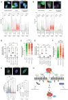
TRIP12’s controller function in DNA repair pathway engagement and choice. (A) TRIP12’s role in Polβ engagement to γH2AX marked chromatin. Representative images show decreased colocalization (white) of Polβ (green) and γH2AX (red) in TRIP12-depleted cells. Violin dot plots show the distribution of copGFP-Polβ, γH2AX, and Polβ/γH2AX colocalized foci in scrambled (SCR) shRNA-expressing or TRIP12-depleted (TRIP12-sh1 and -sh5) unirradiated control (0 Gy) cells and 5 h after 10 Gy irradiation (n = 3 independent experiments with >100 cells each); *P< .05, **P< .01, ***P< .001, and ****P< .0001 as determined by Kruskal–Wallis. (B) Mean colocalized foci/cell values per experiment, pooled in panel (A), and corresponding statistical evaluations (*P< .05 and **P< .01 from ANOVA) are shown. (C) The relative numbers of Polβ colocalized with γH2AX (on a cell-by-cell basis) in irradiated cells are shown with medians and interquartile ranges. (D) Role of TRIP12 ubiquitylation site in Polβ engagement to γH2AX marked chromatin. Representative images show decreased colocalization (white) of the TRIP12 ubiquitylation-mutated copGFP-Polβ(TM/DM) isoform (green) and γH2AX (red). Violin dot plots show the distribution of the copGFP-Polβ isoforms [copGFP-Polβ(WT), copGFP-Polβ(TM), copGFP-Polβ(TM/DM), as indicated], γH2AX, and Polβ/γH2AX colocalized foci in control (0 Gy) cells and 5 h after 10 Gy irradiation (n = 3 independent experiments with >100 cells each); *P< .05, **P< .01, ***P< .001, and ****P< .0001 as determined by the Kruskal–Wallis test. (E) Mean foci/cell values per experiment and corresponding statistical evaluations (*P< .05 from ANOVA) are shown. (F) The relative numbers of Polβ [copGFP-Polβ(WT), copGFP-Polβ(TM), or copGFP-Polβ(TM/DM)] colocalized with γH2AX (on a cell-by-cell basis) are shown with medians and interquartile ranges. (G) Influence of TRIP12 on colocalization of Polβ with 53BP1. CopGFP-Polβ and 53BP1 foci colocalization in control (SCR) and TRIP12 knockdown cells (sh1 and sh5) 5 h after 10 Gy irradiation are shown and compared to unirradiated controls (0 Gy) in violin dot plots illustrating changes in median and distribution (left graph) and as mean foci/cell values from two independent experiments with at least n = 100 cells each (right graph; see Supplementary Fig. S5 for additional data). Asterisks mark P-values (Kruskal–Wallis test, ****P< .0001, **P< .01) assessing the impact of TRIP12 depletion and radiation on these cells. (H) TRIP12 in the governance of repair pathway choice. A graphic is shown that illustrates the current working model of TRIP12 involvement in BER. TRIP12-mediated prevention of the RNF168 promoted histone ubiquitylation extension surrounding DNA lesions restrains DSB signaling in favor of Polβ. Enabled by its ubiquitylation activity on Polβ, TRIP12 exerts its repair traffic control function by assisting BER and deviating DSB repair at the same time. Complex damage sites with clustered lesions can cause DSBs if not repaired appropriately and DSB repair attempts at DSBs residing in such regions may fail due to the presence of such BER-targeted lesions, since they prevent synthesis and nuclease activities. A cellular control mechanism that channels repair proteins to yield the right of way to BER may be therefore relevant in these cases. A two-step activity can be proposed in which TRIP12 engages Polβ at nuclear damage sites but also initiates BER complex disassembly, freeing Polβ from XRCC1 for distinct repair activities and thereafter destines Polβ for proteasome degradation upon repair completion.

Update of

References

    1. Hustedt N, Durocher D The control of DNA repair by the cell cycle. Nat Cell Biol. 2017; 19:1–9. 10.1038/ncb3452. - DOI - PubMed
    1. Clouaire T, Legube G DNA double strand break repair pathway choice: a chromatin based decision?. Nucleus. 2015; 6:107–13. - PMC - PubMed
    1. Fang Q, Inanc B, Schamus S et al. HSP90 regulates DNA repair via the interaction between XRCC1 and DNA polymerase β. Nat Commun. 2014; 5:5513. 10.1038/ncomms6513. - DOI - PMC - PubMed
    1. Kakarougkas A, Jeggo PA DNA DSB repair pathway choice: an orchestrated handover mechanism. Br J Radiol. 2014; 87:20130685. 10.1259/bjr.20130685. - DOI - PMC - PubMed
    1. Kratz A, Kim M, Kelly MR et al. A multi-scale map of protein assemblies in the DNA damage response. Cell Syst. 2023; 14:447–63.e448. 10.1016/j.cels.2023.04.007. - DOI - PMC - PubMed

LinkOut - more resources