. 2025 Jun 20;53(12):gkaf585.
doi: 10.1093/nar/gkaf585.

Structural basis of a dual-function type II-B CRISPR-Cas9

Affiliations

Structural basis of a dual-function type II-B CRISPR-Cas9

Grace N Hibshman et al. Nucleic Acids Res. .

Abstract

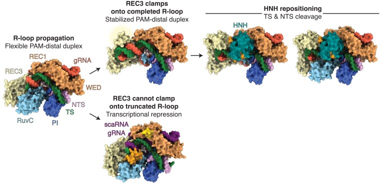
Cas9 from Streptococcus pyogenes (SpCas9) revolutionized genome editing by enabling programmable DNA cleavage guided by an RNA. However, SpCas9 tolerates mismatches in the DNA-RNA duplex, which can lead to deleterious off-target editing. Here, we reveal that Cas9 from Francisella novicida (FnCas9) possesses a unique structural feature-the REC3 clamp-that underlies its intrinsic high-fidelity DNA targeting. Through kinetic and structural analyses, we show that the REC3 clamp forms critical contacts with the PAM-distal region of the R-loop, thereby imposing a novel checkpoint during enzyme activation. Notably, F. novicida encodes a noncanonical small CRISPR-associated RNA (scaRNA) that enables FnCas9 to repress an endogenous bacterial lipoprotein gene, subverting host immune detection. Structures of FnCas9 with scaRNA illustrate how partial R-loop complementarity hinders REC3 clamp docking and prevents cleavage in favor of transcriptional repression. The REC3 clamp is conserved across type II-B CRISPR-Cas9 systems, pointing to a potential path for engineering precise genome editors or developing novel antibacterial strategies. These findings reveal the molecular basis of heightened specificity and virulence enabled by FnCas9, with broad implications for biotechnology and therapeutic development.


Conflict of interest statement

None declared.

Figures

Graphical Abstract
Graphical Abstract
Figure 1.
Figure 1.
Visualization of distinct stages of FnCas9 nuclease activation. (A) FnCas9 domain organization with the REC3 clamp crosshatched. (B) 2.9 Å cryo-EM reconstruction of FnCas9 in a nonproductive state with no RuvC density resolved. Ovals indicate missing cryo-EM density. The corresponding models are depicted to the right of the cryo-EM reconstructions. (C) 2.9 Å cryo-EM reconstruction of FnCas9 in a nonproductive state with RuvC density. (D) 3.0 Å cryo-EM reconstruction of FnCas9 in a productive state with HNH in the active conformation and no RuvC density. (E) 2.6 Å cryo-EM reconstruction of FnCas9 in a productive state with RuvC density. (F) Detailed view of the HNH active site with the cryo-EM map overlaid. (G) Detailed view of the REC3 clamp and HNH L1 interactions with the cryo-EM map overlaid.
Figure 2
Figure 2
The REC3 clamp enhances FnCas9 specificity. (A) Surface representation of FnCas9 in the product state with the nucleotides colored by observed cleavage rate for a single mismatch at that TS position. PM represents a perfect match DNA substrate. Observed cleavage rates were measured for each mismatch via capillary electrophoresis. Red indicates a slower observed cleavage rate, and blue indicates a faster observed cleavage rate. (B) Detailed view of REC3 clamp residues contacting the PAM-distal heteroduplex in the product state conformation. (C) Schematic representation of REC3 contacts with the PAM-distal heteroduplex. Dashed lines represent hydrogen bonds; solid lines represent other contacts; asterisks represent the residues mutated to alanine; red circles represent water molecules. (D) 100 nM of each FnCas9 variant was mixed with 25 nM of each substrate, and the cleaved DNA product was monitored via capillary electrophoresis. Percent cleaved reported after 1 h incubation. Black denotes wild-type FnCas9; purple denotes the FnCas9 clamp mutant. (E, F) FnCas9 was incubated with DNA containing a mismatch at position 16 of the TS for 1 h. The reaction was quenched via vitrification and analyzed via cryo-EM. (E) Thirty percent of the particles (79,858 particles) from this dataset yielded a 2.9 Å cryo-EM reconstruction of FnCas9 in a productive state bound to DNA with a mismatch at position 16. (F) Seventy percent of the particles (128,469 particles) from this dataset comprised a 3.0 Å cryo-EM reconstruction of FnCas9 in a nonproductive state bound to DNA with a mismatch at position 16.
Figure 3.
Figure 3.
The REC3 clamp senses target complementarity distinguishing DNA cleavage from transcriptional repression. (A) Schematic of the F. novicida chromosomal locus consisting of Cas genes, tracrRNA, crRNA, and scaRNA. (B) Schematic of canonical FnCas9 targeting where 20 bp of R-loop form between the gRNA and TS. (C) Detailed view of L1 when the R-loop has propagated to completion and HNH is in the active conformation. (D) Schematic of scaRNA-mediated targeting where 11 bp of complementarity exists prior to encountering mismatches. 2.8 Å cryo-EM reconstruction of scaRNA-mediated targeting by FnCas9 with 14 bp of R-loop formed. (E) Detailed view of L1 in scaRNA-mediated transcriptional repression structure with mismatches shown in red.
Figure 4.
Figure 4.
Type II-B CRISPR–Cas systems contain extended REC3 domains. (A) Detailed view of REC3 clamp residues contacting the PAM-distal heteroduplex colored by conservation score. The location of these residues is highlighted in panels (B) and (C). (B) Surface representation of FnCas9 in the product state where each residue is colored by conservation, where purple represents more conserved residues, and cyan denotes more variable residues. (C) Domain schematic of FnCas9 with the corresponding conservation score for each residue. Residues with a conservation score of 225 or higher are considered highly conserved. (D) Structural comparison of type II CRISPR–Cas9 enzymes, including type II-A SpCas9 (PDB 7S4X), type II-C NmeCas9 (PDB 6JDV), type II-B FnCas9 (this study), PsCas9 (PDB 8UMF), and LpCas9 (AF3). The REC3 domains are labeled for each structure. The REC2 domain is unresolved in the FnCas9 structure as indicated by the circle.
Figure 5.
Figure 5.
Model of type II-B CRISPR–Cas9 nuclease activation. If the R-loop propagates to completion, the REC3 clamp docks onto the PAM-distal heteroduplex prompting HNH repositioning and DNA cleavage. Alternatively, if REC3 cannot clamp due to incomplete R-loop formation, type II-B CRISPR–Cas9 repress transcription without cleavage.

Update of

Similar articles

References

    1. Sorek R, Lawrence CM, Wiedenheft B CRISPR-mediated adaptive immune systems in bacteria and archaea. Annu Rev Biochem. 2013; 82:237–66. 10.1146/annurev-biochem-072911-172315. - DOI - PubMed
    1. Jinek M, Chylinski K, Fonfara I et al. A programmable dual-RNA-guided DNA endonuclease in adaptive bacterial immunity. Science. 2012; 337:816–21. - PMC - PubMed
    1. Mali P, Yang L, Esvelt KM et al. RNA-guided human genome engineering via Cas9. Science. 2013; 339:823–6. - PMC - PubMed
    1. Cong L, Ann Ran F, Cox D et al. Multiplex genome engineering using CRISPR/Cas systems. Science. 2013; 339:819–23. - PMC - PubMed
    1. Fu Y, Foden JA, Khayter C et al. High-frequency off-target mutagenesis induced by CRISPR–Cas nucleases in human cells. Nat Biotechnol. 2013; 31:822–6. - PMC - PubMed

MeSH terms

Supplementary concepts

LinkOut - more resources