. 2025 Sep:85:103744.
doi: 10.1016/j.redox.2025.103744. Epub 2025 Jun 26.

Gaylussacin, a stilbene glycoside, inhibits chronic obstructive pulmonary disease in mice

Affiliations

Gaylussacin, a stilbene glycoside, inhibits chronic obstructive pulmonary disease in mice

Hye-Young Min et al. Redox Biol. 2025 Sep.

Abstract

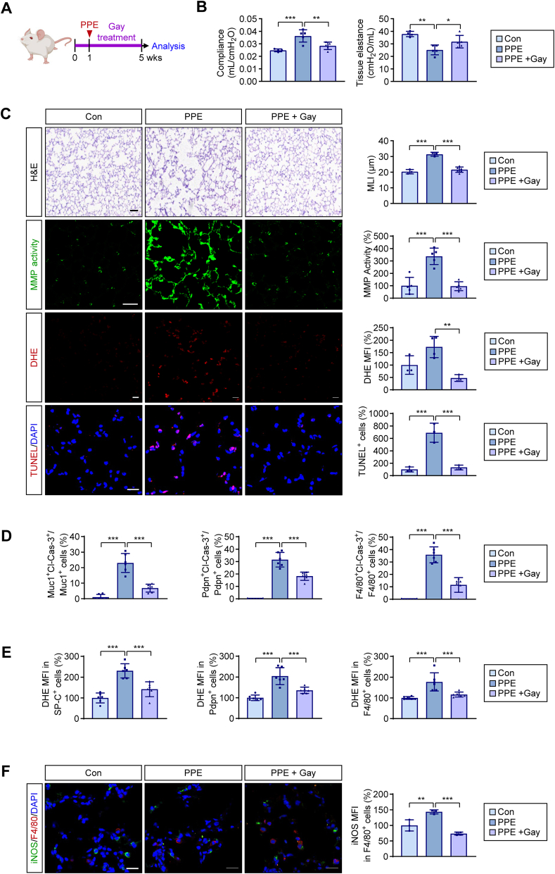
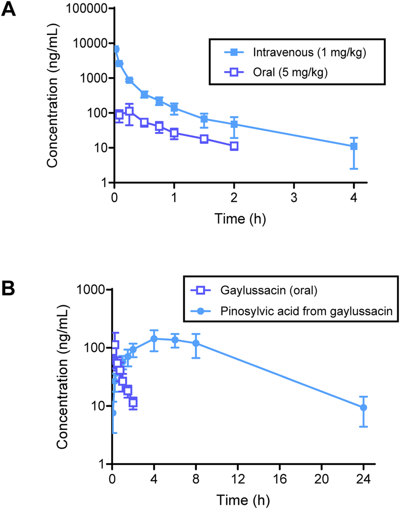
Chronic obstructive pulmonary disease (COPD) is a major cause of human mortality worldwide and is closely associated with chronic inflammation triggered by environmental toxicants such as lead (Pb) and cadmium (Cd). However, the molecular mechanisms linking Pb/Cd exposure to COPD pathogenesis and effective therapeutic strategies remain poorly defined. In this study, we established a mouse model of environmentally induced COPD by exposing mice to Pb/Cd aerosols using a specialized nebulizer system. Pb/Cd exposure led to characteristic COPD-like pathological features, including alveolar damage, mucus hypersecretion, oxidative stress, and apoptosis. Transcriptome analysis of lung tissues revealed upregulation of pro-inflammatory cytokines, chemokines, and lipid metabolism-related genes, with macrophages-particularly those expressing MMP-12-identified as key contributors to pulmonary inflammation. Through a targeted stilbenoid compound screen, we identified gaylussacin as a potent suppressor of Pb/Cd-induced MMP-12 expression in macrophages. Mechanistically, gaylussacin suppressed expression of MMP-12 and inflammatory mediators via activation of SIRT1. In a porcine pancreatic elastase (PPE)-induced emphysema model, oral administration of gaylussacin significantly improved lung function, reduced apoptosis, ROS production, and inflammation. Pharmacokinetic analysis revealed limited oral bioavailability of gaylussacin but efficient conversion to its active metabolite, pinosylvic acid. Toxicological evaluations confirmed negligible toxicity in normal cells derived from various organs and no significant adverse effects in vivo. Collectively, these findings demonstrate that Pb/Cd inhalation promotes COPD pathogenesis through macrophage-driven inflammation mediated by MMP-12 and that gaylussacin mitigates these effects by enhancing SIRT1 activity. This study supports gaylussacin as a promising therapeutic candidate for the treatment of environmentally induced COPD.

Keywords: Chronic obstructive pulmonary disease; Gaylussacin; Heavy metal; Matrix metalloproteinase-12.


Conflict of interest statement

Declaration of competing interest The authors declare that they have no known competing financial interests or personal relationships that could have appeared to influence the work reported in this paper.

Figures

Fig. 1
Fig. 1
Pathogenic alterations with global transcriptomic changes in lungs from chronic lead and cadmium exposure. FVB/N mice inhaled an aqueous solution of lead and cadmium (Pb/Cd) for 3 months. (A) Schematic depicting the experimental schedule. (B, C) Analysis of changes in the architecture (B, mean linear intercept [MLI]), mucus hypersecretion (B, PAS positivity), reactive oxygen species (ROS) production (B, DHE positivity), matrix metalloproteinase (MMP) activity (B, in situ zymography), and cleavage of caspase-3 (C, Cl-Cas-3 expression) in mouse lungs exposed to Pb/Cd (mean ± SD, n = 3 or 4/group). Scale bars: 100 μm (H&E and PAS staining) and 20 μm (cleaved caspase-3, MMP activity, and DHE staining). (D) Immunofluorescence (IF) staining of Cl-Cas-3 expression in Muc1+ AT2 cells, Pdpn+ AT1 cells, and F4/80+ macrophages in the lungs of control or Pb/Cd-treated mice (mean ± SD, n = 6/group). Representative IF images are shown in Supplementary Fig. 1. (E) DAVID analysis revealed significant enrichment of gene ontology (GO, biological process and molecular function) terms in the Pb/Cd-treated group compared with the control group. The list of genes (n = 839) that were significantly upregulated in the Pb/Cd group (fold change >2 and p < 0.05) was used for the DAVID analysis. “Count” represents the number of genes corresponding to each GO term among the list of genes. (F) Real-time PCR analysis assessed the expression regulation of various genes associated with pro- and anti-inflammatory responses, and lipid metabolism, in murine lungs from the Pb/Cd group compared to those of the control group (mean ± SD, n = 3/group). (G) IF staining of F4/80+ macrophage recruitment in the lungs of mice exposed to Pb/Cd (mean ± SD, n = 4/group). Scale bar: 20 μm. (H) IF staining showing an increase in the lipid accumulation (Nile red+) within F4/80+ macrophages in the lungs of mice following chronic Pb/Cd inhalation. Scale bars: 20 μm. (I) Analysis of changes in the architecture (MLI), mucus hypersecretion (PAS positivity), ROS production (DHE positivity), MMP activity (in situ zymography), and Cl-Cas-3 expression in mouse lungs exposed to Pb, Cd, or their combination (mean ± SD, n = 7/group). Representative images are shown in Supplementary Fig. 2. ∗p < 0.05; ∗∗p < 0.01; ∗∗∗p < 0.001, as determined using a two-tailed Student's t-test (B–D, F, G), Mann-Whitney test (D), and one-way ANOVA with Tukey's multiple comparison test (I). Con: control.
Fig. 2
Fig. 2
The identification of matrix metalloproteinase-12 (MMP-12) as a marker for the development of emphysematous lung changes induced by chronic lead and cadmium inhalation and for COPD development in humans. (A) The Venn diagram identifies common genes associated with gene ontology (GO) terms related to proteolysis and collagen. (B) Real-time PCR analysis for the validation of the regulation of the specified gene expression in murine lungs of the Pb/Cd group compared to that of the control group (mean ± SD, n = 3/group). (C) Western blot analysis of the MMP-12 expression in the lungs of control or Pb/Cd-inhaled mice. Densitometric analysis was performed using ImageJ software (mean ± SD, n = 6/group). (D) Immunofluorescence (IF) staining of MMP-12 expression in the lungs of control or Pb/Cd-inhaled mice (mean ± SD, n = 8/group). Scale bars: 20 μm. (E) IF staining of MMP-12 expression in F4/80+ macrophages in the lungs of control or Pb/Cd-inhaled mice (mean ± SD, n = 16 or 18/group). Scale bars: 20 μm. (F) Real-time PCR analysis was used to validate MMP12 expression levels, showing increased regulation in human lungs with emphysematous lesions compared to normal human lungs (mean ± SD, n = 5/group). (G) The violin plot shows differential expression of MMP12 in various cell types in the lungs of patients with COPD in comparison with those of healthy controls, as determined by analysis of a publicly available dataset (GSE136831). (H) Violin plots show a significant increase in MMP12 expression in macrophages, a subpopulation of conventional dendritic cells (cDC2), and monocytes in the lungs of patients with COPD in comparison with those of healthy controls. Wilcoxon statistics and significance levels were added on the top of the violin plots. ∗p < 0.05; ∗∗p < 0.01; ∗∗∗p < 0.001, as determined using a two-tailed Student's t-test (B–D, F) and Welch's t-test (E). Con: control.
Fig. 3
Fig. 3
Association of matrix metalloproteinase-12 (MMP-12) expression in macrophages with the development of pulmonary inflammation and emphysematous lung changes induced by chronic lead and cadmium inhalation. (A) Real-time PCR analysis of Mmp12 expression in MH-S, MLg, and MLE-12 cells that were exposed to a mixture of Pb (5 μM) and Cd (1 μM) for 1, 7, and 30 d (mean ± SD, n = 3/group). (B) Real-time PCR and western blot analyses showing an increase in MMP-12 mRNA (mean ± SD, n = 3/group) and protein expression following combined treatment of MH-S cells with Pb (5 μM) and Cd (1 μM) for 1 d. (C) Western blot analysis showing an increase in pro- and cleaved forms of MMP-12 in the conditioned medium (CM) of MH-S cells treated with vehicle (control) or a mixture of Pb (5 μM) and Cd (1 μM) for 24 h. The Coomassie blue staining of the gel indicates a loading control, ensuring equal protein loading for the conditioned medium. (D) The single-cell RNA sequencing of the lungs from control mice and Pb/Cd-treated mice revealed the distribution of Mmp12 expression across different cell types in the lungs, comparing the case group (mice treated with Pb/Cd for 6 months) to the control group, as shown in feature plots. (E) Violin plots show differential expression of Mmp12 expression in alveolar macrophage and fibroblast between the case and control groups. Wilcoxon statistics and significance levels were added on the top of the violin plots. (F) Feature plots show Mmp12 expression in the subgroups of AT2 cell populations between case and control groups. (G) The violin plot shows Mmp12 expression in AT2 cells between case and control groups. Wilcoxon statistics and significance levels were added on the top of the violin plots. (H) Real-time PCR analysis of the indicated gene expression in Mmp12 expression in tissue-resident alveolar macrophages (TR-AMs) and circulating adult bone marrow–derived monocytes (Mo-AMs) isolated from the lungs of mice exposed to a mixture of Pb and Cd for 3 months (mean ± SD, n = 3/group). ∗p < 0.05; ∗∗p < 0.01; ∗∗∗p < 0.001, as determined using a two-tailed Student's t-test (A, B, H). Con: control.
Fig. 4
Fig. 4
Identification of gaylussacin as an inhibitor of MMP-12 expression with negligible toxicity in vitro. (A) Structures of the seven stilbenoids used in this study. (B) Effect of stilbenoids (10 μM) on Mmp12 expression in MH-S cells (mean ± SD, n = 3/group). Cells were pretreated with various stilbenoids (10 μM) for 1 d and further stimulated with a combination of Pb (5 μM) and Cd (1 μM) for 2 d in the presence of test compounds. (C, D) Effect of stilbenoids (10 μM) on MMP-12 expression in the whole-cell lysates (WCLs, C) and the conditioned medium (CM, D) in MH-S cells (mean ± SD, n = 3/group). Cells were pretreated with test compounds for 2 h and then treated with a combination of Pb (5 μM) and Cd (1 μM) for 24 h in the presence of test compounds. (E) Effect of stilbenoids (10 μM) on the viability of MH-S cells. MH-S cells were treated with various stilbenoids (10 μM) for 2 d. Cell viability was determined using the crystal violet assay (mean ± SD, n = 5/group). (F) Real-time PCR analysis for the expression of Sirt1, Mmp12, and pro-inflammatory genes in MH-S cells that were transiently transfected with empty or SIRT1-expressing vectors and exposed to a mixture of Pb (5 μM) and Cd (1 μM) for 1 d. (G) Effects of gaylussacin on Pb/Cd-mediated regulation of SIRT1 activity and the expression of Mmp12 and pro-inflammatory genes. MH-S cells were treated with gaylussacin (Gay, 10 μM) for 2 h and then exposed to a mixture of Pb (5 μM) and Cd (1 μM) in the absence of presence of gaylussacin for 24 h. p < 0.05; ∗∗p < 0.01; ∗∗∗p < 0.001, as determined using one-way ANOVA with Dunnett's multiple comparison test for comparison with the control group (B, E, F, G). Con: control.
Fig. 5
Fig. 5
Blockade of elastase-induced emphysematous lesions via oral administration of gaylussacin in mice. (A) Schematic depicting the experimental schedule. (B) Effect of gaylussacin (Gay, 40 mg/kg) on elastase (porcine pancreatic elastase, PPE)-induced pulmonary dysfunction. Changes in lung function were monitored using FlexiVent (mean ± SD, n = 4 or 5/group). (C) Effect of gaylussacin (Gay) on elastase-mediated pulmonary damage, MMP activity, ROS production, and apoptotic cell death in murine lungs, as determined via H&E staining, in situ zymography using fluorescein-conjugated DQ-gelatin, DHE staining, and TUNEL staining, respectively. Right. Quantification of structural changes in the airspace, gelatinase activity, DHE positivity, and TUNEL positivity (mean ± SD, n = 3–6/group). Scale bars: 25 μm (H&E and MMP activity); 20 μm (DHE staining); 50 μm (TUNEL staining). (D) Immunofluorescence (IF) staining of cleaved caspase-3 (Cl-Cas-3) expression in Muc1+ AT2 cells, Pdpn+ AT1 cells, and F4/80+ macrophages in the lungs of the indicated group of mice (mean ± SD, n = 6/group). Representative IF images are shown in Supplementary Fig. 4A. (E) IF staining of ROS production (DHE positivity) in SP-C+ AT2 cells, Pdpn+ AT1 cells, and F4/80+ macrophages in the lungs of the indicated group of mice (mean ± SD, n = 6/group). Representative IF images are shown in Supplementary Fig. 4B. (F) Effect of gaylussacin on elastase-mediated iNOS expression in F4/80+ macrophages in the lungs, as determined via IF staining of cryosections of lung tissues. Right. Quantification of iNOS expression levels in F4/80-positive cells compared with the vehicle-treated control (mean ± SD, n = 3 or 4/group). Scale bars: 20 μm. p < 0.05; ∗∗p < 0.01; ∗∗∗p < 0.001, as determined using a one-way ANOVA with Dunnett's multiple comparison test, comparing results with the PPE-treated group. Con: control.
Fig. 6
Fig. 6
Pharmacokinetics of gaylussacin in rats. (A) The plasma concentration-time profiles of gaylussacin after intravenous (1 mg/kg) or oral (5 mg/kg) administration in rats (mean ± SD, n = 4/group). (B) The plasma concentration-time profile of pinosylvic acid after oral administration of gaylussacin (5 mg/kg) (mean ± SD, n = 4/group). The profile of gaylussacin in this analysis is consistent with that observed in (A).
Fig. 7
Fig. 7
Minimal toxic effects of gaylussacin in vitro and in vivo. (A) Gaylussacin showed no significant cytotoxic effects on normal cell lines from various organs. Cells were treated with vehicle (DMSO) or gaylussacin (1, 5, 10, and 50 μM) for 2 d. Cell viability was determined using the crystal violet assay (mean ± SD, n = 4/group). (B) Body weight changes in each treatment group in the animal experiment (mean ± SD, n = 3 or 4/group). (C) Serum ALT and AST levels in vehicle- or gaylussacin (40 mg/kg)- treated mice were measured (mean ± SD, n = 5/group). No significant changes observed, based on one-way ANOVA with Dunnett's multiple comparison test for cell viability and body weight (A, B) and a two-tailed Student's t-test for ALT and AST levels (C). Con: control. Gay: gaylussacin.

References

    1. Barnes P.J., et al. Chronic obstructive pulmonary disease. Nat. Rev. Dis. Primers. Dec 3 2015;1 doi: 10.1038/nrdp.2015.76. - DOI - PubMed
    1. G. B. D. C. o. D. Collaborators Global burden of 288 causes of death and life expectancy decomposition in 204 countries and territories and 811 subnational locations, 1990-2021: a systematic analysis for the global burden of disease study 2021. Lancet. May 18 2024;403(10440):2100–2132. doi: 10.1016/S0140-6736(24)00367-2. - DOI - PMC - PubMed
    1. Horio Y., et al. Protective effect of Galectin-9 in murine model of lung emphysema: involvement of neutrophil migration and MMP-9 production. PLoS One. 2017;12(7) doi: 10.1371/journal.pone.0180742. - DOI - PMC - PubMed
    1. Tachkov K., Kamusheva M., Pencheva V., Mitov K. Evaluation of the economic and social burden of chronic obstructive pulmonary disease (COPD) Biotechnol. Biotechnol. Equip. 2017/07/04 2017;31(4):855–861. doi: 10.1080/13102818.2017.1335616. - DOI
    1. Tchounwou P.B., Yedjou C.G., Patlolla A.K., Sutton D.J. Heavy metal toxicity and the environment. Experientia Suppl. 2012;101:133–164. doi: 10.1007/978-3-7643-8340-4_6. - DOI - PMC - PubMed

MeSH terms

LinkOut - more resources