

White piedra: fungal extracellular matrix formation and its importance in pathogenesis. An ultrastructural study
- PMID: 40614550
- PMCID: PMC12270794
- DOI: 10.1016/j.abd.2025.501140
White piedra: fungal extracellular matrix formation and its importance in pathogenesis. An ultrastructural study
Abstract
Background: White piedra is a disease caused by some species of the genus Trichosporon. A case of white piedra was investigated, whose molecular examination identified Cutaneotrichosporon (Trichosporon) debeurmannianum as the causative agent.
Methods: Scanning electron microscopy (SEM) was used to examine the affected hairs, as well as the fungal colony of C. debeurmannianum obtained from the hairs. For comparative purposes, a colony of Trichosporon mucoides obtained from a mycotheque was also examined.
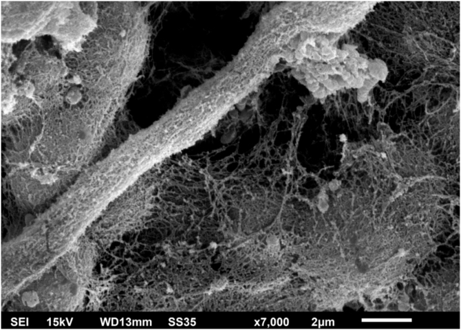
Results: Examination of the affected hairs using SEM easily demonstrates nodules on the hair shaft with a predominance of rounded yeast-like structures, adhered to each other by a cementing substance. Examination of the C. debeurmannianum colony demonstrates significant adhesion between the fungal cells by a reticular extracellular matrix. Examination of the T. mucoides colony obtained from a mycotheque demonstrates a small production of fibrillar substance between the blastoconidia.
Discussion: Examination of the colony obtained from the piedra showed significant formation of extracellular matrix, adhering to and covering the fungal structures, forming a biofilm. This matrix must correspond to the cementing substance described in the condition.
Conclusion: The synthesis of the extracellular matrix must be crucial in the formation of white piedra nodules.
Keywords: Extracellular matrix; Scanning electron microscopy; White piedra.
Copyright © 2025 Sociedade Brasileira de Dermatologia. Published by Elsevier España, S.L.U. All rights reserved.
Conflict of interest statement
Conflicts of interest None declared.
Figures


References
-
- Diniz LM, De Souza Filho JB. Estudo de 15 casos de piedra branca observados na Grande Vitória (Espírito Santo ‒ Brasil) durante cinco anos. An Bras Dermatol. 2005;80:49–52.
-
- Magalhães A.R., Mondino S.S., Silva Md, Nishikawa M.M. Morphological and biochemical characterization of the aetiological agents of white piedra. Mem Inst Oswaldo Cruz. 2008;103:786–790. - PubMed
-
- Roselino A.M., Seixas A.B., Thomazini J.A., Maffei C.M. An outbreak of scalp white piedra in a Brazilian children day care. Rev Inst Med Trop Sao Paulo. 2008;50:307–309. - PubMed
Publication types
MeSH terms
LinkOut - more resources
Full Text Sources
