. 2025 Jul 4;16(1):6067.
doi: 10.1038/s41467-025-60248-9.

Immunocompetent cell targeting by food-additive titanium dioxide

Affiliations

Immunocompetent cell targeting by food-additive titanium dioxide

John W Wills et al. Nat Commun. .

Abstract

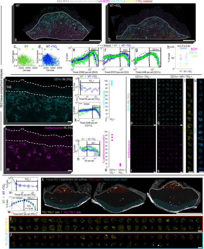
Food-grade titanium dioxide (fgTiO2) is a bio-persistent particle under intense regulatory scrutiny. Yet paradoxically, the only known cell reservoirs for fgTiO2 are graveyard intestinal pigment cells which are metabolically and immunologically quiescent. Here we identify immunocompetent cell targets of fgTiO2 in humans, most notably in the subepithelial dome region of intestinal Peyer's patches. Using multimodal microscopies with single-particle detection and per-cell / vesicle image analysis we achieve correlative dosimetry, quantitatively recapitulating human cellular exposures in the ileum of mice fed a fgTiO2-containing diet. Epithelial microfold cells selectively funnel fgTiO2 into LysoMac and LysoDC cells with ensuing accumulation. Notwithstanding, proximity extension analyses for 92 protein targets reveal no measureable perturbation of cell signalling pathways. When chased with oral ΔaroA-Salmonella, pro-inflammatory signalling is confirmed, but no augmentation by fgTiO2 is revealed despite marked same-cell loading. Interestingly, Salmonella causes the fgTiO2-recipient cells to migrate within the patch and, sporadically, to be identified in the lamina propria, thereby fully recreating the intestinal tissue distribution of fgTiO2 in humans. Immunocompetent cells that accumulate fgTiO2 in vivo are now identified and we demonstrate a mouse model that finally enables human-relevant risk assessments of ingested, bio-persistent (nano)particles.


Conflict of interest statement

Competing interests: J.W. states that after completing the presented work as University of Cambridge Herchel-Smith Fellow, he subsequently undertook employment with the pharmaceutical company, GlaxoSmithKline. A.D., J.R., M.M., S.R., H.D.S., R.E.H., A.F., A.B.D.S., C.A.P.B., S.M., Å.V.K., J.D.S., N.C.R., D.O., R.J., P.M., A.P.B., P.R. and J.J.P. declare no competing interests.

Figures

Fig. 1
Fig. 1. Titanium dioxide in human intestinal tissues.
a Tilescanned image acquired by confocal reflectance microscopy. Reflectant foci were detected along the follicle base consistent with previously-reported mineral particle-containing pigment cells. Of note, similar reflectant foci were also present apically in the immuno-active, subepithelial dome tissue-region, and away from the patch in the (c) lamina propria of the regular villous mucosa (VM). Translucent red circle-markers are placed on the reflectant foci to aid visualisation. bde, Correlative SEM/EDX analyses performed on the same tissue section shown in (a/c). The pigment cell region is shown in (b) where X-ray signal for both Al (attributable to aluminosilicates) and Ti (attributable to titanium dioxide) was found. EDX analyses in the (d) villous mucosa and (e) subepithelial dome regions showed reflectant foci were attributable to Ti. b, d, e In all instances the elemental data for Ti were a near-exact match for the reflectant foci, whereas (b) areas with Al signal alone did not reflect. The image-data presented in (ae) are representative of independent repeats in n = 2 samples. f Reflectance microscopical analyses of nine, randomly-drawn human samples (six frozen, three formalin-fixed paraffin embedded (FFPE)) showed reflectant foci consistent with TiO2 in the subepithelial dome of every sample. Scale bars: (a) = 50 μm; (b, d, e) = 10 μm; (cf) = 50 μm with 10 μm insets.
Fig. 2
Fig. 2. A mouse feeding study demonstrates fgTiO2 specificity for Peyer’s patches.
a Single-cell analysis of a tilescanned ileal tissue section. As in humans (Fig. 1a/f), fgTiO2 loaded into the subepithelial dome (SED) and there was evidence of precursor pigment cell formation along the follicle base. Beyond the follicle, two positive cells (a, insets) were found in the villous mucosa. In contrast to the human findings (Fig. 1c) both were caused by luminal fgTiO2 trapped in the invaginated space of goblet cells. bd To investigate the sensitivity of fgTiO2 detection by confocal reflectance microscopy, a tissue-region containing a single, reflectant foci was milled out under correlative SEM. e-i Transfer of the lamella to a transmission electron microscope enabled imaging and X-ray (EDX) and electron diffraction analysis. A (g, h) single particle of fgTiO2 with an (i) anatase diffraction pattern (i.e., as added to the diet) was confirmed responsible for the single reflectant foci observed. j Caecal patches (adjacent at the top of the colon) were almost completely devoid of fgTiO2 (n = 4 animals). k Immunofluorescence microscopy showed fgTiO2 access to Peyer’s patches was via GP2-positive microfold (M) cells. l Single-cell analysis of a longitudinal ileal section consistently showed the close association between an M-cell rich, follicle-associated epithelium and fgTiO2 uptake. m, n Guided by CD11c and CD3 labelling for phagocytic mononuclear cells and T-lymphocytes (respectively) image-sets were collected from the SED, germinal centre (GC) T-cell zone (TCZ) or overlying villous mucosa (VM) tissue regions (n = 4 mice per diet-group). Some fgTiO2 signal was observed in the GC and TCZ regions but the majority was in the SED. In keeping with (a), and in contrast to the human findings (Fig. 1c), in all four mice, no uptake was seen in the villous mucosa demonstrating specificity for M-cell mediated, Peyer’s patch targeting. Scale bars: a = 500 μm with insets 5 μm; b = 50 μm; c = 10 μm; d = 5 μm; e = 500 nm; g = 200 nm; h = 20 nm; i = 10 1/nm; j = 500 μm with insets 10 μm; k = 10 μm; l, m = 500 μm.
Fig. 3
Fig. 3. Establishing correlative human-mouse dosimetry.
al Confocal reflectance microscopy showing the range of cellular loading in the subepithelial dome tissue region of (af), six randomly-drawn human and (g–l) six randomly-drawn mouse samples. After acquisition, the image-fields per specimen were manually laid out in order of lowest-to-highest fgTiO2 SED accumulation to visually present the wide variation in fgTiO2 cellular loading. Translucent red circle-markers were placed on thresholded reflectant foci to aid visualisation. mp Quantitative analysis of the image-data shown in (al) (n = 6 mice / n = 6 humans). m Thresholded reflectance per unit tissue area (i.e., amount of fgTiO2 per unit tissue area). n Thresholded reflectance per cell (i.e., fgTiO2 dose per cell). o, Number of thresholded reflectant foci per cell (i.e., number of fgTiO2-loaded vesicles (TLV) per cell). p Thresholded reflectance signal per foci (i.e., fgTiO2 dose per TLV). Statistical comparison of the distributions is presented in Supplementary Fig. 6 (two-sided Wilcoxon rank-sum analysis). None of the sets of measured fgTiO2 distributions were found to be entirely unique to either species. mp By all of the quantitative measures established, feeding a murine diet supplemented with 0.0625% (w/w) fgTiO2 for eighteen weeks provides significant overlap with measured, real-world human exposures. Scale bars: al = 50 μm with insets 10 μm; n = 20 μm; p = 5 μm.
Fig. 4
Fig. 4. fgTiO2 selectively targets PD-L1+ LysoMac and LysoDC cells.
ah In situ, single-cell analysis of key immune-cell subtypes of the mouse subepithelial dome. a, b Example images immunofluorescently labelled for B220, CD3 and CD11c (i.e., identifying B-lymphocytes, T-lymphocytes and phagocytic mononuclear cells, respectively) from wild-type (WT) mice fed (a) without (WT) or (b) with (WT + TiO2) fgTiO2 dietary supplementation (0.0625% w/w of diet). fgTiO2 was measured from thresholded reflectance images. Translucent red circle-markers are placed on reflectant foci to aid visualisation. c, d Flow cytometry-type plots showing cell area/cell aspect ratio, (eg) immunofluorescence distributions and (h) cell counts for B220 + , CD11c+ and CD3+ cells (n = 3 animals). All measures remained similar regardless of fgTiO2 feeding (cell count comparison (h), P = ≥ 0.26, two-sided unpaired samples T-tests). il fgTiO2-recipient cells of the subepithelial dome were CD11c+ in ~ 95% of cases. mo These cells also exhibited a unique autofluorescent signature as previously described for subepithelial dome LysoMac and LysoDC cells. o Again, this was true for almost all fgTiO2 + /CD11c+ cells and, equally, these autofluorescent cells were similarly present in (n) controls without fgTiO2 feeding (further data shown, Supplementary Fig. 8). pu Montaged CD11c + /fgTiO2+ or CD11c + /fgTiO2- cell populations from the SEDs of mice fed the fgTiO2 supplemented diet visualised in terms of (r, s) autofluorescence signature. fgTiO2 is seen to both selectively and specifically target the autofluorescent cells and (t, u) the diversity of CD4 and MHCII expression confirms that both LysoMac and LysoDCs take up fgTiO2. vz fgTiO2+ cells of the SED expressed the immunotolerance marker programmed death ligand 1 (PD-L1) whereas fgTiO2+ pigment cells at the base of the Peyer’s patch tended to lose PD-L1 expression (y versus (z), respectively). w PD-L1 expression in the Peyer’s patches was not significantly perturbed by feeding the fgTiO2-supplemented diet (P = 0.34, z = −0.96, two-sided Wilcoxon rank-sum analysis, n = 6 animals). The image-data presented in (i, m, pu, xz) were collected from SED tissue sections from n = 3 mice. Scale bars: a, b = 50 μm; i, m, pu = 10 μm; x = 500 μm; y, z = 10 μm.
Fig. 5
Fig. 5. fgTiO2-Salmonella interactions.
Mouse Study 2 used a 16-week fgTiO2 feeding period before switching all animals to a normal diet (i.e., without fgTiO2-supplementation) then orally inoculating half with attenuated, ΔaroA-Salmonella. Tissues were harvested +3 or +28 days after infection. ac Z-stack maximum projections showing immunofluorescent labelling for flagellin (FliC) to detect the ΔaroA-Salmonella in the Peyer’s patch SED region. Marked accumulation of both fgTiO2 and ΔaroA-Salmonella was observed in autofluorescent, phagocytic mononuclear cells at the +3 timepoint. d Faecal ELISA studies confirmed a Salmonella-specific IgA response at the +28 timepoint (bars represent medians with interquartile range error-bars, n = 3–6 samples per treatment group). eg Protein expression analyses of ileal tissue digests by proximity extension assay (OLINK mouse exploratory panel). e Comparison of ileal tissues taken from wild-type (WT) mice unexposed to fgTiO2 but with (WT +Sal) or without (WT) ΔaroA-Salmonella infection confirmed significant increases key cytokines/chemokines associated with a Th1 immune response (P ≤ 0.05, two-sided Wilcoxon rank-sum test, n = 5–6 tissues per group). f Inflammation-associated protein expression analyses of ileal tissue digests. At each timepoint, no significant differences in protein expression were observed between the WT and WT +TiO2 (P ≥ 0.53, two-sided Wilcoxon rank-sum test, n = 3 animals per group) or WT +Sal and WT +TiO2 +Sal groups (P ≥ 0.10, two-sided Wilcoxon rank-sum test, n = 6 animals per group). Full dataset is shown in Supplementary Fig. 12. g Peyer’s patch-focussed inflammation-associated protein expression analyses using carefully-excised patch digests in mice treated with either ΔaroA-Salmonella alone (WT +Sal) or with fgTiO2 and ΔaroA-Salmonella (WT +TiO2 +Sal) at the +3 or +28 timepoints. At each timepoint, no significant differences were observed between the two groups (P ≥ 0.20, two-sided Wilcoxon rank-sum test, n = 6 animals per group) demonstrating no interaction of fgTiO2 and ΔaroA-Salmonella despite (ac) heavy loading into the same cells (full dataset shown in Supplementary Fig. 13). OLINK protein abbreviations are defined in Supplementary Table 1. All p-values are available for download at the BioStudies database under accession number S-BSST875. Scale bars: ac = 10 μm.
Fig. 6
Fig. 6. Cellular dosimetry of fgTiO2 across timepoints and Salmonella-induced migration.
a, b, k Confocal reflectance micrographs showing the subepithelial dome (SED) tissue region (n = 3 animals per group). After the 16-week fgTiO2 feeding period, all animals were switched to a normal diet (without fgTiO2 supplementation) and half were orally dosed with ΔaroA-Salmonella before tissue harvest at +3 or +28-day timepoints. a, b In the animals fed the fgTiO2-supplemented diet without Salmonella exposure (WT +TiO2), fgTiO2-loaded cells were present in the SEDs at both +3 and +28 timepoints. cj To gain insights into cellular dosimetry changes across the two timepoints, image analysis of Peyer’s patch transverse sections was used to measure the number of reflectant foci per-cell (i.e., the fgTiO2-loaded vesicle (TLV) count per-cell) and the amount of reflectance per foci (i.e., equivalent to the fgTiO2 dose per-vesicle). d, e At the +28 timepoint, the TLV count per-cell was elevated with a new population of cells with TLV counts >13 forming in the mid-to-base regions of the follicle. f, g Randomly sampling and montaging the reflectance (RL) information in cells from each of the two timepoints visually demonstrates this, showing (g) more heavily-loaded cells at the +28 timepoint. h Moving from cell to vesicle information, in contrast, the amount of reflected light per individual foci remained near-identical across the two timepoints and this was again borne out visually when (i, j) individual TLVs were randomly montaged. Collectively, the results suggest that once fgTiO2-loaded cells move below the SED, there is a process of vesicular inheritance by certain cells over time, eventually yielding heavily-loaded pigment cells at the follicle base–as is well-described in humans (Fig. 1). k Challenge with ΔaroA-Salmonella (WT +TiO2 +Sal) markedly changed this picture (n = 4 animals per group). At the +28 timepoint, SEDs were almost completely devoid of both fgTiO2 and autofluorescence-positive cells. Out of four SEDs imaged, only one reflectant foci was detectable (indicated by arrow) showing marked migration of fgTiO2 from the SED tissue compartment after ΔaroA-Salmonella exposure. Scale bars: a, b, k = 50 μm, c, d = 250 μm, f, g, i, j = 10 μm.
Fig. 7
Fig. 7. Ileal distribution of fgTiO2 following Salmonella challenge.
a, b Single-cell image analysis of tilescanned confocal reflectance images collected from transverse sections of mouse ileal tissue from the fgTiO2-exposed, ΔaroA-Salmonella-infected treatment group (+28-day timepoint, n = 3 animals; FR denotes follicular region with no follicle-associated epithelium). b As in Fig. 6, the subepithelial dome (SED) regions were largely devoid of fgTiO2 with the majority of the positive cells located at the follicle base in the pigment cell zone or in the interfollicular regions (fgTiO2-positive cells in the Peyer's patches are displayed in red). b In contrast to all previous data collected without ΔaroA-Salmonella exposure, fgTiO2-positive cells were now also present in the villous mucosa (VM) (displayed in white). c Montaging positive events from villous mucosa regions showed that particle loading was low compared to Peyer’s patch cells with the majority containing single reflectant foci. d High resolution imaging of optically cross-sectioned villi confirmed reflectant foci deep in the lamina propria in the same manner as observed in humans (Fig. 1c). e The majority of the fgTiO2-positive events in the villous mucosa also displayed the same autofluorescence signature as was observed in the particle-recipient cells of the SED. The image-data presented in c-e were collected from ileal sections from n = 3 mice. Scale bars: a, b = 250 μm; ce = 20 μm.

References

    1. Riedle, S. et al. Pro-inflammatory adjuvant properties of pigment-grade titanium dioxide particles are augmented by a genotype that potentiates interleukin 1β processing. Part. Fibre Toxicol.14, 51 (2017). - PMC - PubMed
    1. Ashwood, P., Thompson, R. P. & Powell, J. J. Fine particles that adsorb lipopolysaccharide via bridging calcium cations may mimic bacterial pathogenicity towards cells. Exp. Biol. Med.232, 107–117 (2007). - PubMed
    1. Grand View Research. Titanium dioxide market size, share and trends analysis report by grade (anatase, rutile), by production process (sulfate, chloride), by application (paints and coatings, plastics), by region, and segment forecasts, 2021−2028. https://www.grandviewresearch.com/industry-analysis/titanium-dioxide-ind... (2025).
    1. Safety assessment of titanium dioxide (E171) as a food additive. EFSA J. 19, e06585 (2021). - PMC - PubMed
    1. European Medicines Agency. Final feedback from European Medicines Agency (EMA) to the EU Commission request to evaluate the impact of the removal of titanium dioxide from the list of authorised food additives on medicinal products. https://www.ema.europa.eu/en/documents/report/final-feedback-european-me... (2025).