. 2025 Jul 4;15(1):23953.
doi: 10.1038/s41598-025-09221-6.

Homocysteine induces ferroptosis in renal tubular epithelial cells via β-catenin/GPX4 signaling pathway

Affiliations

Homocysteine induces ferroptosis in renal tubular epithelial cells via β-catenin/GPX4 signaling pathway

Yanping Lei et al. Sci Rep. .

Abstract

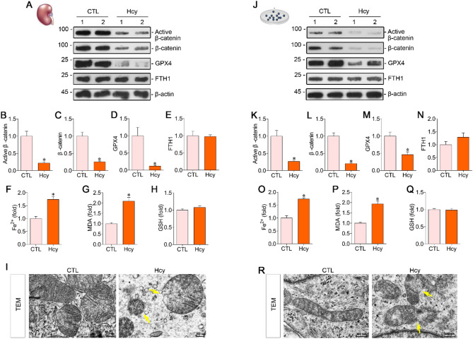
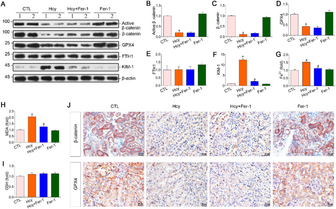
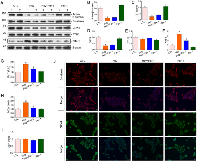
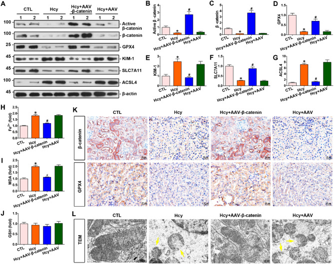
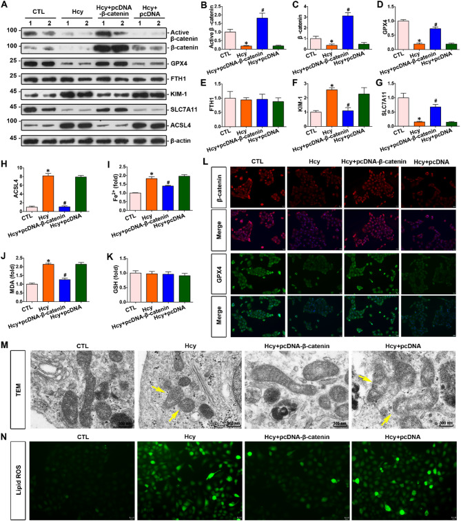
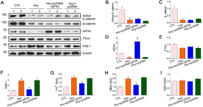
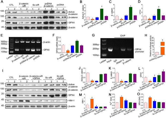
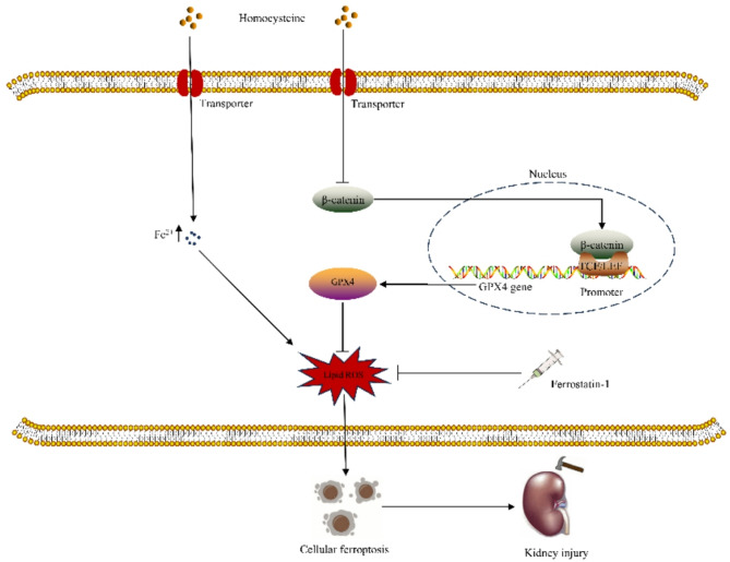
Hyperhomocysteinemia can cause severe damage to kidney. Ferroptosis represents a critical mechanism in the initiation and development of kidney disorders. We focus on the β-catenin/GPX4 signaling pathway to explore how homocysteine influences ferroptosis regulation in renal tubular epithelial cells. C57BL/6J mice were administered drinking water with high level of homocysteine to establish a hyperhomocysteinemia model. In the cell experiments, HKC-8 cells were exposed to homocysteine for a duration of 12 h. Active β-catenin, β-catenin, GPX4, FTH1, and KIM-1 were detected using Western blotting; Biochemical assays were conducted to measure lipid ROS, Fe2+, and GSH; GPX4 and β-catenin were detected through immunohistochemistry and immunofluorescence techniques; Mitochondrial damage was examined using transmission electron microscopy; ChIP analysis, coupled with dual-luciferase reporter gene assays, was employed to investigate the relationship between β-catenin protein and GPX4 gene promoter. Our findings revealed that homocysteine disrupted β-catenin signaling, inhibited GPX4 expression in renal tubular epithelial cells, subsequently promoted ferroptosis. Overexpression of β-catenin or GPX4 inhibited ferroptosis induced by homocysteine, and β-catenin regulated GPX4 expression in renal tubular epithelial cells. Further assays demonstrated that GPX4 acted as a target gene of β-catenin. In conclusion, homocysteine elicits ferroptosis in renal tubular epithelial cells by disrupting β-catenin signaling and inhibiting its target gene, GPX4.

Keywords: Ferroptosis; GPX4; Hyperhomocysteinemia; Renal tubular epithelial cells; Β-catenin.


Conflict of interest statement

Declarations. Competing interests: The authors declare no competing interests. Ethics approval: This study protocol was reviewed and approved by the Animal Care and Use Committee of University of South China (Ethics approval number: USC2023XS087).

Figures

Fig. 1
Fig. 1
Homocysteine impaired β-catenin signaling and promoted ferroptosis in renal tubular epithelial cells. The hyperhomocysteinemia model was established by administering drinking water containing 1.8 g/L homocysteine to the mice. In the in vitro experiments, HKC-8 cells were treated with homocysteine at a concentration of 500 µmol/L. (A) Western blotting was employed to evaluate the expression levels of active β-catenin, β-catenin, GPX4, and FTH1 in the kidneys of mice. (BE) Quantitative analysis was conducted to determine the levels of active β-catenin, β-catenin, GPX4, and FTH1 in the kidneys of mice. (FH) Biochemical assays were performed to measure the levels of Fe2+, MDA, and GSH in mouse kidney tissues. (I) The morphology of mitochondria in renal tubular epithelial cells was examined using transmission electron microscopy. Yellow arrows highlighted mitochondrial damage, including the rupture of the mitochondrial membrane and the reduction of mitochondrial cristae. Scale bar, 300 nm. (J) Western blotting was employed to evaluate the expression levels of active β-catenin, β-catenin, GPX4, and FTH1 in HKC-8. (KN) Quantitative analysis was conducted to determine the levels of active β-catenin, β-catenin, GPX4, and FTH1 in HKC-8. (OQ) Biochemical assays were performed to measure the levels of Fe2+, MDA, and GSH in HKC-8. (R) Mitochondrial morphology in HKC-8 cells was examined using electron microscopy. Yellow arrows highlighted mitochondrial damage, including the rupture of the mitochondrial membrane and the reduction of mitochondrial cristae. Scale bar, 300 nm. *P < 0.05 vs. the controls (n = 6). Hcy, homocysteine; TEM, transmission electron microscopy.
Fig. 2
Fig. 2
Fer-1 alleviated renal ferroptosis triggered by hyperhomocysteinemia without altering β-catenin and GPX4 levels. The ferroptosis inhibitor Fer-1 (2 mg/kg/day, i.p.) was administered to mice with hyperhomocysteinemia, and its therapeutic efficacy was evaluated. (A) Western blotting was used to assess the expression of active β-catenin, β-catenin, GPX4, FTH1, and KIM-1 in the kidneys of mice from indicated groups. (BF) Quantitative analysis of the levels of active β-catenin, β-catenin, GPX4, FTH1, and KIM-1 in Figure (A) was performed. (GI) Biochemical determination of the levels of Fe2+, MDA, and GSH in the kidney tissues of mice from indicated groups was conducted. (J) Immunohistochemical analysis of β-catenin and GPX4 in the renal tissues was performed. Scale bar, 20 μm. *P < 0.05 vs. the controls; #P < 0.05 vs. hyperhomocysteinemia mice (n = 6). Hcy, homocysteine.
Fig. 3
Fig. 3
Fer-1 selectively attenuated homocysteine-induced ferroptosis in HKC-8 cells without affecting β-catenin signaling and the levels of GPX4. The ferroptosis inhibitor Fer-1 (10 µmol/L) was used to treat homocysteine-stimulated HKC-8 cells, and its therapeutic effect was evaluated. (A) Western blotting was used to assess the expression of active β-catenin, β-catenin, GPX4, FTH1, and KIM-1 in HKC-8 cells from indicated groups. (BF) Quantitative analysis of the levels of active β-catenin, β-catenin, GPX4, FTH1, and KIM-1 in Figure (A) was performed. (GI) Biochemical determination of the levels of Fe2+, MDA, and GSH in HKC-8 cells from indicated groups was conducted. (J) Immunofluorescence was employed for the detection of β-catenin and GPX4 in HKC-8 cells. Scale bar, 25 μm. *P < 0.05 vs. the controls; #P < 0.05 vs. homocysteine stimulation alone (n = 6). Hcy, homocysteine.
Fig. 4
Fig. 4
Restoration of β-catenin signaling enhanced GPX4 expression and inhibited ferroptosis induced by hyperhomocysteinemia in the kidneys. Adeno-associated virus expressing β-catenin was used to infect mice with hyperhomocysteinemia, and the effect of β-catenin overexpression on renal tubules was analyzed. (A) Western blotting was used to assess the expression of active β-catenin, β-catenin, GPX4, KIM-1, SLC7A11, and ACSL4 in the kidneys of mice from indicated groups. (BG) Quantitative analysis of the levels of active β-catenin, β-catenin, GPX4, KIM-1, SLC7A11, and ACSL4 in Figure (A) was performed. (HJ) Biochemical determination of the levels of Fe2+, MDA, and GSH in the kidney tissues of mice from indicated groups was conducted. (K) Immunohistochemical analysis of β-catenin and GPX4 in the renal tissues was performed. Scale bar, 20 μm. (L) Mitochondrial morphology in renal tubular epithelial cells was observed using electron microscopy. Yellow arrows highlighted mitochondrial damage, including mitochondrial membrane rupture, reduction of mitochondrial cristae and mitochondrial shrinkage. Scale bar, 300 nm. *P < 0.05 vs. the controls; #P < 0.05 vs. hyperhomocysteinemia mice (n = 6). Hcy, homocysteine; AAV, adeno-associated virus; TEM, transmission electron microscopy.
Fig. 5
Fig. 5
Upregulation of β-catenin signaling enhanced GPX4 expression and markedly suppressed ferroptosis in HKC-8 cells induced by elevated homocysteine levels. The β-catenin plasmid was transfected into homocysteine-treated HKC-8 cells, and the effect of β-catenin overexpression on the renal tubular epithelium was analyzed. (A) Western blotting was used to assess the expression of active β-catenin, β-catenin, GPX4, FTH1, KIM-1, SLC7A11, and ACSL4 in HKC-8 cells from indicated groups. (BH) Quantitative analysis of the levels of active β-catenin, β-catenin, GPX4, FTH1, KIM-1, SLC7A11, and ACSL4 in Figure (A) was performed. (IK) Biochemical determination of the levels of Fe2+, MDA, and GSH in HKC-8 cells from indicated groups was conducted. (L) Immunofluorescence was employed for the detection of β-catenin and GPX4 in cells. Scale bar, 25 μm. (M) Mitochondrial morphology in HKC-8 cells was examined using transmission electron microscopy. Yellow arrows highlighted mitochondrial damage, including mitochondrial membrane rupture, reduction of mitochondrial cristae and mitochondrial shrinkage. Scale bar, 300 nm. (N) Lipid ROS levels were assessed using the fluorescent probe BODIPY581/591 C11 in each group. Scale bar, 25 μm. *P < 0.05 vs. the controls; #P < 0.05 vs. homocysteine stimulation alone (n = 6). Hcy, homocysteine; pcDNA, pcDNA3.1; TEM, transmission electron microscopy.
Fig. 6
Fig. 6
Upregulation of GPX4 expression is capable of inhibiting homocysteine-induced ferroptosis in renal tubular epithelial cells without affecting β-catenin expression levels. The GPX4 plasmid was transfected into homocysteine-treated HKC-8 cells, and the effect of GPX4 overexpression on HKC-8 cells was analyzed. (A) Western blotting was used to assess the expression of active β-catenin, β-catenin, GPX4, FTH1, and KIM-1 in HKC-8 cells from indicated groups. (BF) Quantitative analysis of the levels of active β-catenin, β-catenin, GPX4, FTH1, and KIM-1 in Figure (A) was performed. (GI) Biochemical determination of the levels of Fe2+, MDA, and GSH in HKC-8 from indicated groups was conducted. *P < 0.05 vs. the controls; #P < 0.05 vs. homocysteine stimulation alone (n = 6). Hcy, homocysteine; pcDNA, pcDNA3.1.
Fig. 7
Fig. 7
As a target gene of β-catenin, enhanced GPX4 expression counteracted the ferroptosis induced by downregulated β-catenin signaling. β-catenin siRNA and pcDNA-β-catenin were transfected into HKC-8 cells to assess the effect of β-catenin on GPX4 expression. (A) Western blotting was used to assess the expression of active β-catenin, β-catenin, and GPX4 in HKC-8 cells from indicated groups. (BD) Quantitative analysis of the levels of active β-catenin, β-catenin, and GPX4 in Figure (A) was performed. *P < 0.05 vs. the controls (n = 6). (E) The mRNA levels of GPX4 in each group were analyzed using RT-PCR. (F) The mRNA levels of GPX4 in figure (E) were quantitatively analyzed. *P < 0.05 vs. the controls (n = 6). (G) ChIP analysis of the interaction between β-catenin and the promoter region of the GPX4 gene. (H) Dual-luciferase reporter gene assay was used to analyze the regulatory effect of β-catenin on GPX4. *P < 0.05 vs. the controls (n = 4). β-catenin siRNA and GPX4 plasmid were co-transfected into HKC-8 cells to assess their interaction. (I) Western blotting was used to assess the expression of active β-catenin, β-catenin, GPX4, and KIM-1 in HKC-8 cells from indicated groups. (JM) Quantitative analysis of the levels of active β-catenin, β-catenin, GPX4, and KIM-1 in Figure (I) was performed. (NO) Biochemical determination of the levels of Fe2+ and lipid MDA in HKC-8 cells from indicated groups was conducted. *P < 0.05 vs. the controls; #P < 0.05 vs. β-catenin siRNA transfection (n = 6). Sc-siR, scramble siRNA; β-catenin siR, β-catenin siRNA; pcDNA, pcDNA3.1.
Fig. 8
Fig. 8
A graphical abstract of the mechanism underlying homocysteine-induced ferroptosis in renal tubular epithelial cells.

Similar articles

References

    1. Yang, B. et al. Prevalence of hyperhomocysteinemia in china: a systematic review and meta-analysis. Nutrients. 7, 74–90 (2014). - PMC - PubMed
    1. Wu, D. F. & Yin, R. X. Homocysteine, hyperhomocysteinemia, and H-type hypertension. Eur. J. Prev. Cardiol.31, 1092–1103 (2024). - PubMed
    1. Zhang, M. et al. Hyperhomocysteinemia exacerbates ischemia-reperfusion injury-induced acute kidney injury by mediating oxidative stress, DNA damage, JNK pathway, and apoptosis. Open. Life Sci.16, 537–543 (2021). - PMC - PubMed
    1. Pushpakumar, S., Kundu, S. & Sen, U. Hydrogen sulfide protects hyperhomocysteinemia-Induced renal damage by modulation of Caveolin and eNOS interaction. Sci. Rep.9, 2223 (2019). - PMC - PubMed
    1. Angelini, A. et al. The link between homocysteine, folic acid and vitamin B12 in chronic kidney disease. G Ital. Nefrol. 38, 2021–vol2024 (2021). - PubMed

MeSH terms

Substances

LinkOut - more resources