. 2025 Jul 4;20(1):80.
doi: 10.1186/s13062-025-00667-2.

CAF-derived exosomal miR-196b-5p after androgen deprivation therapy promotes epithelial-mesenchymal transition in prostate cancer cells through HOXC8/NF-κB signaling pathway

Affiliations

CAF-derived exosomal miR-196b-5p after androgen deprivation therapy promotes epithelial-mesenchymal transition in prostate cancer cells through HOXC8/NF-κB signaling pathway

Xiaodong Song et al. Biol Direct. .

Abstract

Background: Cancer-associated fibroblasts (CAFs) have been reported to play a significant role in the development and metastasis of various tumors; however, research on their role in promoting prostate cancer (PCa) metastasis under castration conditions remains unclear.

Methods: In this study, we utilized quantitative reverse transcription polymerase chain reaction (qRT-PCR) to detect the expression differences of microRNA-196b-5p (miR-196b-5p) in the exosomes secreted by CAFs before and after castration. We further characterized the transcriptional regulatory landscape through RNA sequencing combined with bioinformatics databases. In vitro and in vivo experiments were conducted to determine the role of miR-196b-5p in promoting tumor migration and metastasis. The dual-luciferase reporter assay, RT-PCR analysis, and Western blot analysis confirmed that miR-196b-5p targets HOXC8 in prostate cancer. Additionally, transwell assays and Western blot analysis were performed to elucidate the role and specific mechanisms of HOXC8 in tumor metastasis.

Results: By analyzing the expression differences of miRNAs in the exosomes secreted by CAFs before and after castration, along with relevant data from databases, we found that miR-196b-5p is highly secreted by CAFs after castration. miR-196b-5p promotes the migration and metastasis of prostate cancer cells. Subsequently, through RNA sequencing analysis and experimental validation, we determined that miR-196b-5p targets HOXC8. This interaction activates the NF-κB pathway, leading to the upregulation of epithelial-mesenchymal transition (EMT)-related protein expression, thereby driving the metastasis of prostate cancer.

Conclusions: Our study elucidates a specific mechanism by which CAF-derived exosomes promote prostate cancer metastasis via miR-196b-5p regulation, contributing to the identification of therapeutic targets for managing tumor metastasis following castration.

Keywords: Cancer-associated fibroblasts; Exosome; HOXC8; NF-κB; Prostate cancer; miR-196b-5p.


Conflict of interest statement

Declarations. Ethics approval and consent to participate: Not applicable. Consent for publication: Not applicable. Competing interests: The authors declare no competing interests.

Figures

Fig. 1
Fig. 1
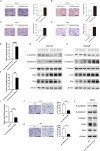
Identification of miR-196b-5p expression levels in primary fibroblasts and prostate cell lines. (A) Immunofluorescence staining of α-SMA and FAP in primary normal prostate fibroblasts, primary prostate CAFs, and hTERT PF179T CAFs (scale bar = 100 μm). (B) The expression levels of miR-196b-5p in primary normal prostate fibroblasts, primary prostate CAFs, and hTERT PF179T CAFs measured by RT-PCR. (C) The expression levels of miR-196b-5p in the normal prostate epithelial cell line RWPE-1 and various prostate cancer cell lines measured by RT-PCR. Data are presented as mean ± SD, representing triplicate measurements. (Student’s t-test, ***P < 0.001)
Fig. 2
Fig. 2
Exosomes transfer miR-196b-5p from CAFs to PCa cells. (A) Expression of miR-196b-5p in exosomes from DHT-treated CAFs and matched ETOH-treated CAFs measured by RT-PCR. (B) Expression of miR-146a-5p in LNCaP and DU145 cells measured by RT-PCR at 24 h after treating with exosomes (25 µg/mL) derived from DHT-treated CAFs or ETOH-treated CAFs. (C) CAFs were transiently transfected with Cy3-labeled miR-control or miR-196b-5p and co-cultured with DU145 cells for 24 h following GW4869 treatment. Fluorescence microscopy was used to observe green signals (β-actin) and red signals (Cy3) in DU145 cells (scale bar = 50 μm). (D) Exosomes were isolated from the supernatant of CAFs transfected with Cy3-labeled miR-196b-5p or miR-control, and exosomes (25 µg/mL) were incubated with DU145 cells for 24 h. Fluorescence microscopy was used to detect green signals (β-actin) and red signals (Cy3) in DU145 cells (scale bar = 50 μm). Data are presented as mean ± SD, representing triplicate measurements. (Student’s t-test, **P < 0.01, ***P < 0.001, ****P < 0.0001)
Fig. 3
Fig. 3
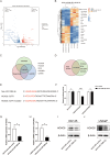
Expression landscape of miR-196b-5p across multiple prostate cancer datasets. (A) Differential expression of miR-196b-5p between tumor and normal samples from the TCGA-PRAD database. (B) Analysis of differential expression of miR-196b in tumor and normal samples from the GSE64333 dataset. (C) Differential expression of miR-196b between CRPC and HSPC samples from the GSE70770 dataset. (D) Comparative differential expression analysis of miR-196b between tumor samples at stages T3–T4 and T2 based on the GSE70770 dataset
Fig. 4
Fig. 4
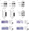
miR-196b-5p promotes cell migration and invasion in PCa cells via activating EMT. (A) and (B) Transwell assays performed to assess the effect of miR-196b-5p on migration (A) and invasion (B) in DU145 cells (scale bars = 25 μm). (C) and (D) Transwell assays performed to assess the effect of miR-196b-5p on migration (C) and invasion (D) in LNCaP cells (scale bars = 25 μm). (E) and (F) Efficiency of miR-196b-5p overexpression in DU145 and LNCaP cells was measured by RT-PCR. (G) and (H) Western blot utilized to assess the effect of miR-196b -5p on EMT marker protein in DU145 and LNCaP cells, respectively. Each set of three lanes in the figure represents three biological replicates. (I) Knockdown efficiency of miR-196b-5p in PC3 cells by miR-196b-5p inhibitor measured using RT-PCR. (J) and (K) Transwell assays performed to assess the effect of miR-196b-5p inhibitor on migration and invasion, respectively (scale bars = 25 μm). (L) Western blot analysis used to detect EMT marker protein in PC3 cells. Data are presented as mean ± SD, representing triplicate measurements. (Student’s t-test, *P < 0.05, **P < 0.01, ***P < 0.001, ****P < 0.0001)
Fig. 5
Fig. 5
HOXC8 is a direct target of miR-196b-5p in PCa cells. (A) Volcano plot of DEGs. (B) Heatmap of DEGs. (C) miR-196b-5p target genes predicted by the MIRWalk, TargetScan, and miRDB databases. (D) Intersection of significantly downregulated DEGs with predicted miR-196b-5p target genes. (E) Predicted target sequence of miR-196b-5p in the 3′-UTR of HOXC8, along with the construction of wild-type and mutant HOXC8 3′-UTR luciferase reporter constructs in HEK-293T cells. (F) The relative luciferase activities were measured after co-transfection of miR-NC mimics or miR-196b-5p mimics and a luciferase vector encoding NC, wild-type, or mutant HOXC8 3′-UTR region. (G) and (H) HOXC8 mRNA levels in DU145 and LNCaP cells transfected with lenti-miR-196b-5p. (I) and (J) HOXC8 protein levels transfected with lenti-miR-196b-5p in DU145 and LNCaP cells. Data are presented as mean ± SD, representing triplicate measurements. (Student’s t-test, *P < 0.05, **P < 0.01, ****P < 0.0001)
Fig. 6
Fig. 6
HOXC8 inhibits EMT in PCa cells. (A) and (B) Western blot analysis to assess the efficiency of HOXC8 overexpression in DU145 and LNCaP cells, respectively. (C) and (D) RT-PCR analysis to assess the efficiency of HOXC8 overexpression in DU145 and LNCaP cells, respectively. (E) and (F) Western blot assessment of EMT marker protein in DU145 and LNCaP cells transfected by HOXC8-control or HOXC8-OE. (G) and (H) Evaluation of the effects of HOXC8 on migration (G) and invasion (H) in DU145 cells by Transwell assays (scale bars = 25 μm). (I) and (J) Evaluation of the effects of HOXC8 on migration (I) and invasion (J) in LNCaP cells by Transwell assays (scale bars = 25 μm). Data are presented as mean ± SD, representing triplicate measurements. (Student’s t-test, *P < 0.05, **P < 0.01, ***P < 0.001, ****P < 0.0001)
Fig. 7
Fig. 7
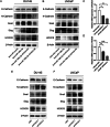
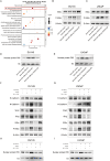
miR-196b-5p promotes EMT in PCa cells by targeting HOXC8. (A) and (B) Western blot analysis to assess the protein levels of E-cadherin, N-cadherin, Snail, Slug, and HOXC8 in DU145 and LNCaP cells transfected with lenti-miR-control, lenti-miR-196b-5p, lenti-miR-196b-5p HOXC8-control, or lenti-miR-196b-5p HOXC8-OE. (C) and (D) Relative expression levels of HOXC8 mRNA in DU145 and LNCaP cells transfected with siHOXC8. (E) and (F) Western blot analysis to assess the protein levels of E-cadherin, N-cadherin, Snail, Slug, and HOXC8 in DU145 and LNCaP cells following transfected with siHOXC8
Fig. 8
Fig. 8
miR-196b-5p promotes metastasis of PCa cells in vivo. (A) Bioluminescence images of BALB/c nude mice that injected with DU145 luciferase cells treated with either miR-196b-5p mimics or miR-NC mimics. The color scale bar depicts the photon flux emitted from the mice. (B) Tumor foci in lungs of BALB/c nude mice after injecting DU145 cells treated with either miR-196b-5p mimics or miR-NC mimics. (C) Immunohistochemical staining of HOXC8 in tumors from mice after injecting DU145 cells treated with either miR-196b-5p mimics or miR-NC mimics. (Student’s t-test, *P < 0.05, **P < 0.01)
Fig. 9
Fig. 9
miR-196b-5p exhibits its functions by activating NF-κB signaling pathway in PCa cells (A) KEGG enrichment analysis of DEGs. (B-E) Western blot analysis of IKBα, p-IKBα, and nuclear protein P65 levels in DU145 and LNCaP cells transfected with lenti-miR-control, lenti-miR-196b-5p, lenti-miR-196b-5p HOXC8-control, or lenti-miR-196b-5p HOXC8-OE. (F-I) Western blot analysis of E-cadherin, N-cadherin, Snail, Slug, IKBα, p-IKBα, and nuclear protein P65 levels in PCa cells treated with NF-κB inhibitor after transfection with lenti-miR-control, lenti-miR-196b-5p, lenti-miR-196b-5p HOXC8-control, or lenti-miR-196b-5p HOXC8-OE
Fig. 10
Fig. 10
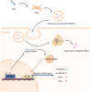
The flowchart of this study, created with BioRender.com

Similar articles

References

    1. Kuo PH, Morris MJ, Hesterman J, Kendi AT, Rahbar K, Wei XX, Fang B, Adra N, Garje R, Michalski JM, et al. Quantitative 68Ga-PSMA-11 PET and clinical outcomes in metastatic Castration-resistant prostate Cancer following 177Lu-PSMA-617 (VISION Trial). Radiology. 2024;312(2):e233460. - PMC - PubMed
    1. Sung H, Ferlay J, Siegel RL, Laversanne M, Soerjomataram I, Jemal A, Bray F. Global Cancer statistics 2020: GLOBOCAN estimates of incidence and mortality worldwide for 36 cancers in 185 countries. Cancer J Clin. 2021;71(3):209–49. - PubMed
    1. Gillessen S, Attard G, Beer TM, Beltran H, Bjartell A, Bossi A, Briganti A, Bristow RG, Chi KN, Clarke N et al. Management of Patients with Advanced Prostate Cancer: Report of the Advanced Prostate Cancer Consensus Conference 2019. European Urology 2020, 77(4):508–547. - PubMed
    1. Wang H, Li N, Liu Q, Guo J, Pan Q, Cheng B, Xu J, Dong B, Yang G, Yang B et al. Antiandrogen treatment induces stromal cell reprogramming to promote castration resistance in prostate cancer. Cancer Cell 2023, 41(7). - PubMed
    1. Sahai E, Astsaturov I, Cukierman E, DeNardo DG, Egeblad M, Evans RM, Fearon D, Greten FR, Hingorani SR, Hunter T, et al. A framework for advancing our Understanding of cancer-associated fibroblasts. Nat Rev Cancer. 2020;20(3):174–86. - PMC - PubMed

MeSH terms

LinkOut - more resources