

Altered endothelial mitochondrial Opa1-related fusion in mouse accelerates age-associated vascular and kidney damage
- PMID: 40616272
- PMCID: PMC12227657
- DOI: 10.14814/phy2.70451
Altered endothelial mitochondrial Opa1-related fusion in mouse accelerates age-associated vascular and kidney damage
Abstract
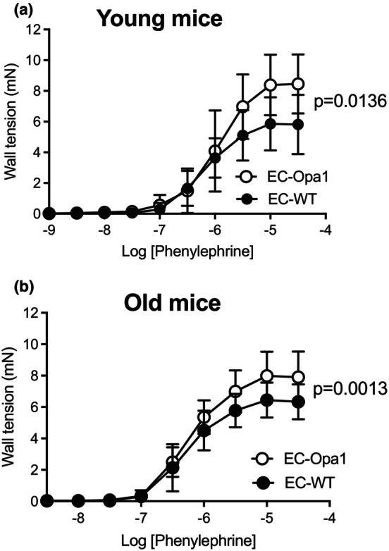
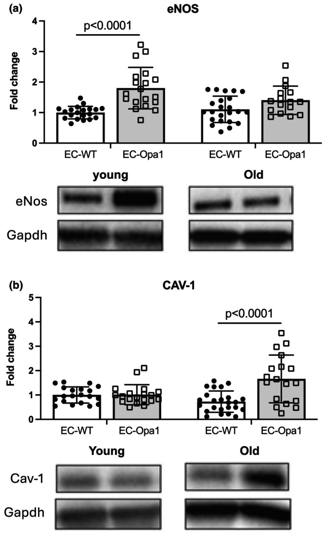
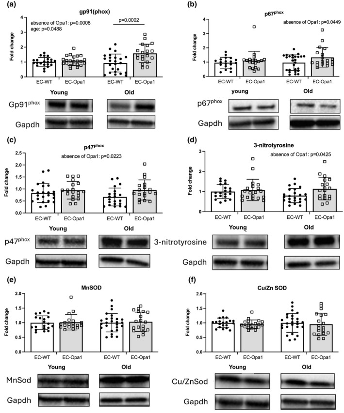
Cardiovascular diseases are the major cause of death worldwide, and their frequency increases with age in association with kidney damage. As a reduction in fusion protein optic atrophy type 1 (Opa1) level in endothelial cells (ECs) decreases the vascular response to flow and increases oxidative stress in perfused kidneys, we hypothesized that reduced Opa1 expression contributes to vascular aging. We used male and female mice with ECs specific Opa1 knock-out (EC-Opa1), and littermate wild-type (EC-WT) mice aged 6 (young) and 20 months (old). Mesenteric resistance arteries (MRA) and kidneys were collected for vascular reactivity and western-blot analysis. In old EC-Opa1 mice, blood urea was greater than in EC-WT mice, and MRA showed reduced endothelium-dependent relaxation. In kidneys, the mitochondria fission protein fission-1 (Fis-1) and the peroxisome proliferator-activated receptor gamma coactivator-1 alpha (Pgc-1α) were increased in old EC-Opa1 mice. The level of caveolin-1 expression was greater in old EC-Opa1 mice. Moreover, in kidneys from EC-Opa1 old mice, NADPH-oxidase subunit gp91 expression was greater than in age-matched EC-WT mice. Thus, reduced mitochondrial fusion in mouse ECs altered mesenteric vascular reactivity and increased markers of oxidative stress in aging kidneys. Thus, Opa1 might protect the vascular tree in target organs such as the kidney.
Keywords: aging; arteries; endothelial cell; kidney; mitochondrial fusion.
© 2025 The Author(s). Physiological Reports published by Wiley Periodicals LLC on behalf of The Physiological Society and the American Physiological Society.
Figures


References
-
- Borri, M. , Jacobs, M. E. , Carmeliet, P. , Rabelink, T. J. , & Dumas, S. J. (2025). Endothelial dysfunction in the aging kidney. American Journal of Physiology. Renal Physiology, 328, F542–F562. - PubMed
-
- Cesareo, M. , Giannini, C. , DI Marino, M. , Aloe, G. , Martucci, A. , Aiello, F. , Cusumano, A. , Mancino, R. , Ricci, F. , Sorge, R. P. , & Nucci, C. (2022). Optical coherence tomography angiography in the multimodal assessment of the retinal posterior pole in autosomal dominant optic atrophy. Acta Ophthalmologica, 100, e798–e806. - PubMed
-
- Chehaitly, A. , Guihot, A. L. , Proux, C. , Grimaud, L. , Aurriere, J. , Legouriellec, B. , Rivron, J. , Vessieres, E. , Tetaud, C. , Zorzano, A. , Procaccio, V. , Joubaud, F. , Reynier, P. , Lenaers, G. , Loufrani, L. , & Henrion, D. (2022). Altered mitochondrial Opa1‐related fusion in mouse promotes endothelial cell dysfunction and atherosclerosis. Antioxidants (Basel), 11, 1078. - PMC - PubMed
-
- Cipolat, S. , Rudka, T. , Hartmann, D. , Costa, V. , Serneels, L. , Craessaerts, K. , Metzger, K. , Frezza, C. , Annaert, W. , D'adamio, L. , Derks, C. , Dejaegere, T. , Pellegrini, L. , D'hooge, R. , Scorrano, L. , & DE Strooper, B. (2006). Mitochondrial rhomboid PARL regulates cytochrome c release during apoptosis via OPA1‐dependent cristae remodeling. Cell, 126, 163–175. - PubMed
MeSH terms
Substances
LinkOut - more resources
Full Text Sources
Medical
Research Materials
