

Impact on the microstructure of deep gray matter in unvaccinated patients after moderate-to-severe COVID-19: insights from MRI T1 mapping
- PMID: 40616705
- PMCID: PMC12228859
- DOI: 10.1186/s41747-025-00598-7
Impact on the microstructure of deep gray matter in unvaccinated patients after moderate-to-severe COVID-19: insights from MRI T1 mapping
Abstract
Background: To determine changes in quantitative T1 relaxation times (qT1) in deep gray matter in patients recovered from coronavirus disease 2019 (COVID-19).
Methods: Unvaccinated COVID-19 participants ≥ 3 months after seropositivity and age- and sex-matched controls were examined using 3-T magnetic resonance imaging. Bilateral measures of thalamus, pallidum, putamen, caudate and accumbens nuclei, and hippocampus were extracted from qT1 maps after automated segmentation. Baseline characteristics and results of tests assessing neurological functions (standardized exam), ability to smell (4-Item Pocket Smell Test), depression (Beck Depression Inventory-II), sleepiness (Epworth Sleepiness Scale), sleep quality (Pittsburgh Sleep Quality Index), health-related quality of life (EQ-5D), and cognitive performance (Montreal Cognitive Assessment) were evaluated.
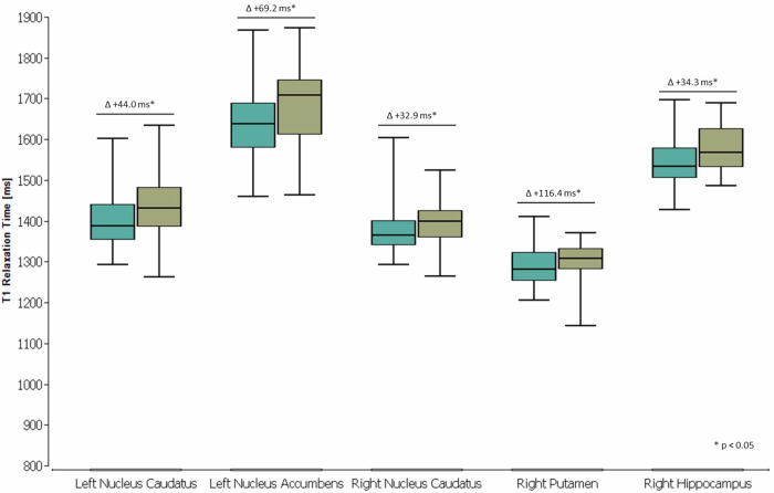
Results: One hundred forty-five subjects (median age, 46 years; 73 females) were included (11/2020-12/2021): 69 recovered after COVID-19 and 76 controls (age, p = 0.532; sex, p = 0.799), without significant differences in qT1 values overall (all p-values > 0.050). Subgroup analysis of participants aged ≥ 40 (age, p = 0.675; sex, p = 0.447) revealed higher qT1 values in previously hospitalized COVID-19 subjects (23/69) compared to controls (47/76) in left and right caudate nuclei (p = 0.009; p = 0.027), left accumbens nucleus (p = 0.017), right putamen (p = 0.041), and right hippocampus (p = 0.020). No correlations were found with macroscopic imaging findings, pre-existing conditions, time since COVID-19 diagnosis, inpatient treatment duration, or test results.
Conclusion: T1 mapping revealed microstructural changes in striatal and hippocampal regions of unvaccinated individuals aged ≥ 40 who recovered from moderate-to-severe COVID-19 during the pre-Omicron era.
Relevance statement: This study elucidates brain involvement following severe acute respiratory syndrome coronavirus 2 (SARS-CoV-2) infection, underscoring the need for further longitudinal analyses to assess the potential reversibility, stability or deterioration of these findings.
Key points: We hypothesized altered T1 relaxation times in deep gray matter after COVID-19. Unvaccinated participants ≥ 40 years exhibited higher striatal, hippocampal qT1 after moderate-to-severe COVID-19. No qT1 correlations were found with hospitalization duration, pre-existing conditions, or neuro-(psycho)logical tests.
Keywords: Brain mapping; COVID-19; Gray matter; Magnetic resonance imaging; SARS-CoV-2.
© 2025. The Author(s).
Conflict of interest statement
Declarations. Ethics approval and consent to participate: The study was approved by the Ethics Committee of the Faculty of Medicine at Goethe University Frankfurt, Germany (20-838) and registered at the German Clinical Trials Register (DRKS00023880). The study was conducted in accordance with the ethical guidelines for research involving human subjects, according to the Declaration of Helsinki. Guarantor: The scientific guarantor of this publication is Prof Dr Elke Hattingen. Consent for publication: Each participant signed an informed consent form for research. Competing interests: The authors declare that they have no competing interests.
Figures

References
MeSH terms
LinkOut - more resources
Full Text Sources
Medical
Miscellaneous