. 2025 Jul 1:85:103749.
doi: 10.1016/j.redox.2025.103749. Online ahead of print.

Ascorbic acid deficiency promotes metabolic remodeling and pulmonary fibrosis that leads to respiratory failure in Sod1 and Akr1a double-knockout mice

Affiliations

Ascorbic acid deficiency promotes metabolic remodeling and pulmonary fibrosis that leads to respiratory failure in Sod1 and Akr1a double-knockout mice

Tsukasa Osaki et al. Redox Biol. .

Abstract

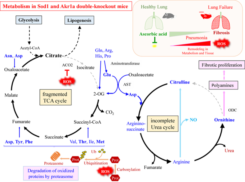
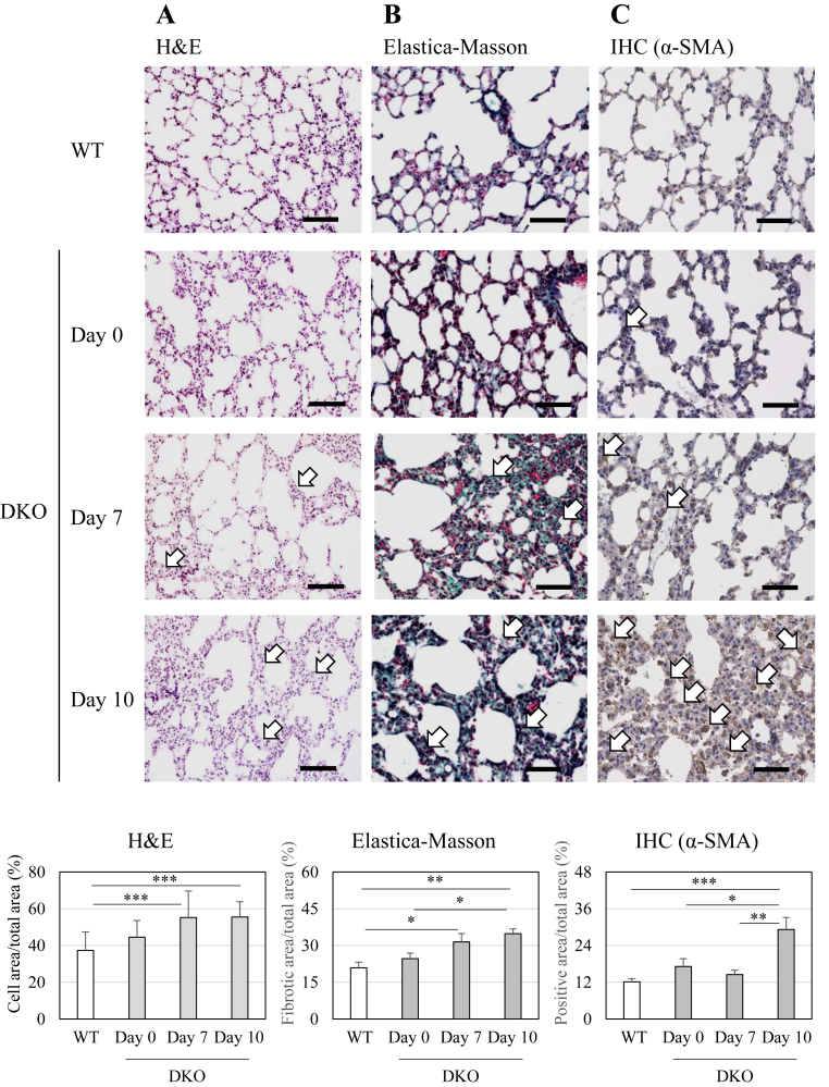
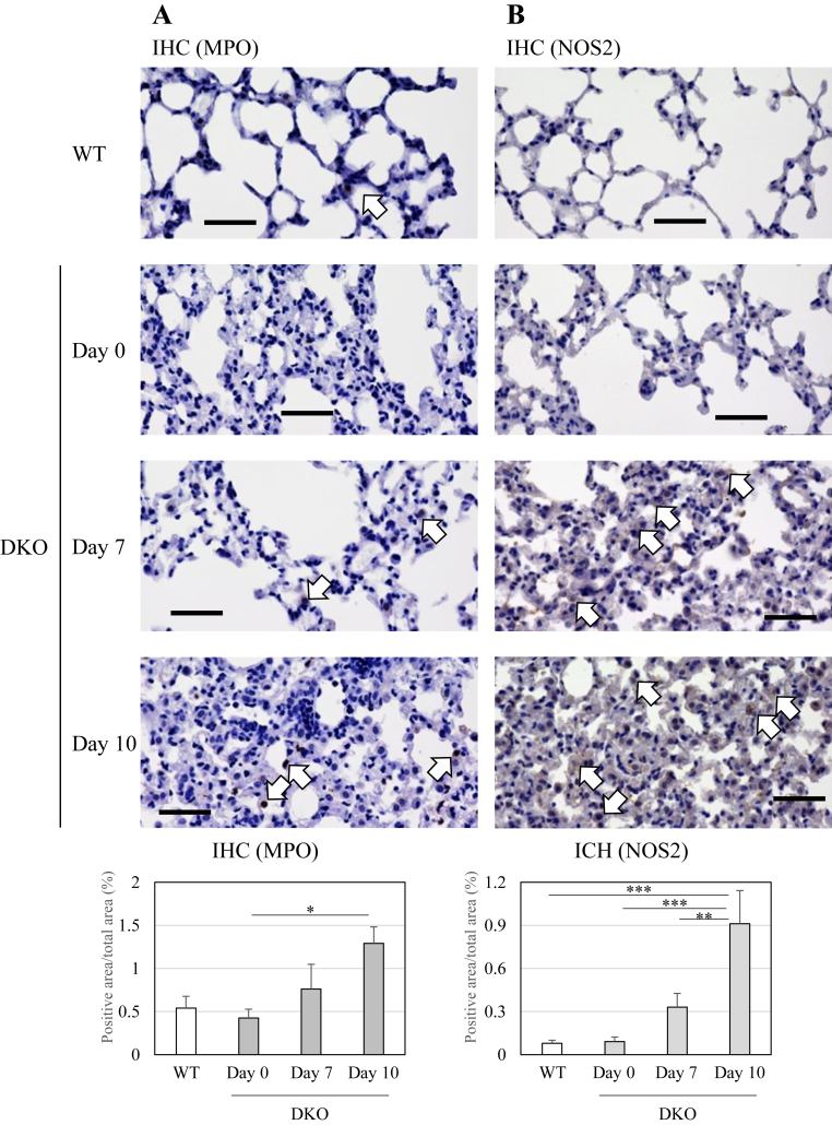
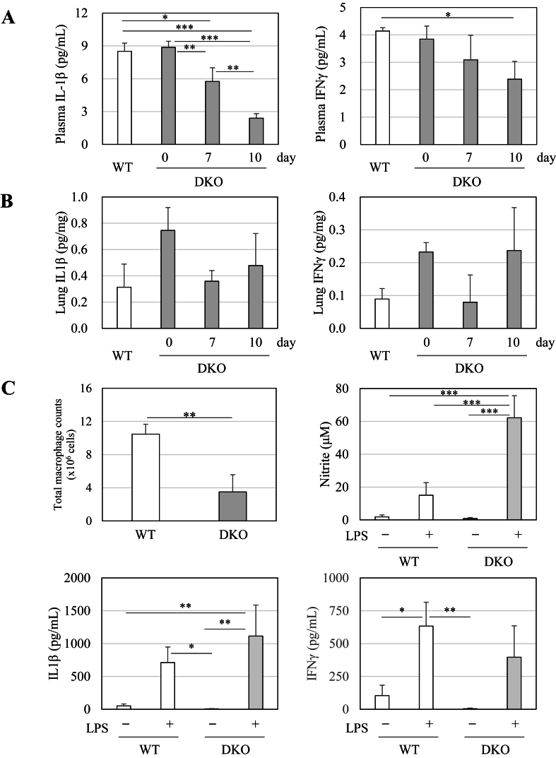
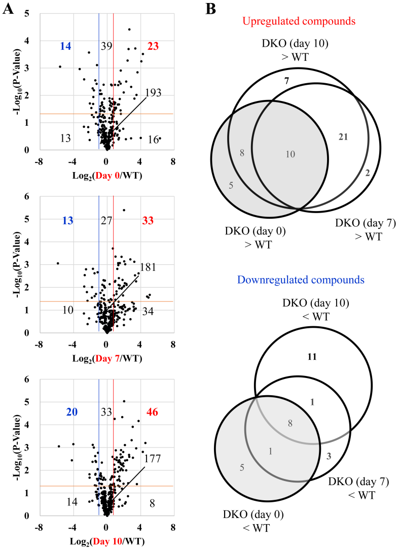
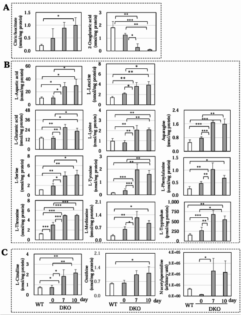
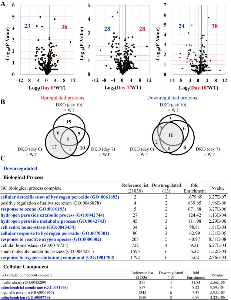
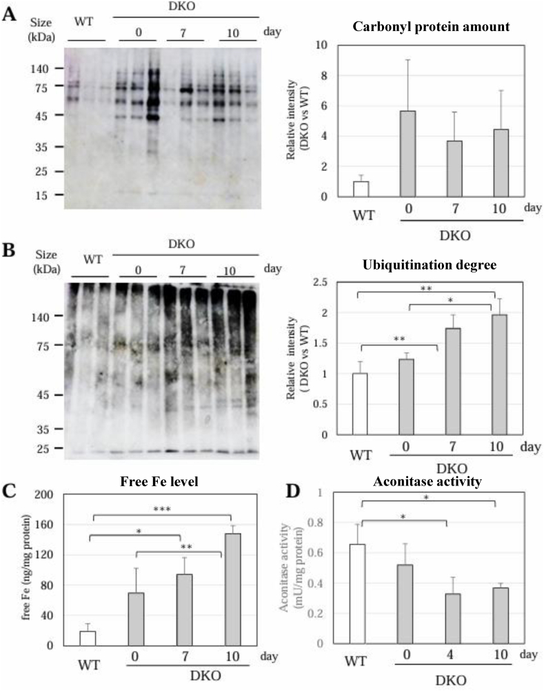
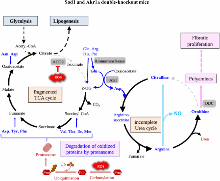
We recently reported that mice with a double knockout (DKO) of Sod1 encoding superoxide dismutase 1 (SOD1) and Akr1a encoding aldehyde reductase survived more than one year when supplemented with ascorbic acid (Asc) (1.5 mg/ml in drinking water), and that the withdrawal of Asc resulted in premature death in only two weeks due to oxidative damage-associated pneumonia. SOD1 is known to disable the radical electrons of superoxide, which suppresses the subsequent formation of highly reactive oxygen species (ROS). Akr1a encodes aldehyde reductase, which catalyzes the biosynthesis of Asc, which is a strong nutritional antioxidant. In this study, we sought to gain insight into the metabolic basis for the progression of respiratory failure in the DKO mice. Pathological examinations have revealed pulmonary damage and the progression of fibrosis caused by an elevation in pulmonary cell death in these mice. Metabolite analyses have shown that substrate compounds catabolized in the tricarboxylic acid cycle are shifted from carbohydrates to amino acids, which leads to polyamine synthesis. While proteins involved in cell polarization, adhesion, and transport are increased in the lungs, showing trends similar to those of activated leukocytes, antioxidative enzymes were characteristically decreased in the lungs. Carbonyl proteins were originally high in the DKO mice but did not increase following Asc withdrawal, which was likely caused by stimulation of the degradation of oxidized proteins through the ubiquitin-proteasome system. It is conceivable that the oxidative insult due to Asc insufficiency under Sod1 deficiency causes protein oxidation followed by degradation, which fuels the tricarboxylic acid cycle. Remodeling the metabolic pathways for amino acid use increases polyamine synthesis, which could stimulate pulmonary fibrosis and lead to respiratory failure.

Keywords: Pneumonia; Proteomics; Superoxide; Tricarboxylic acid cycle; Urea cycle.


Conflict of interest statement

Declaration of competing interest We declare no conflicts of interest between the authors or with any institution in relation to the content of this article.

Figures

Image 1
Graphical abstract
Fig. 1
Fig. 1
Histological evaluation of the lung tissue of WT mice and DKO mice following Asc withdrawal Mice were euthanized at the indicated time points. Lung sections (5 μm in thickness) were subjected to H&E staining (A), Elastica-Masson staining (B), and immunohistochemical staining with anti-α-smooth muscle actin (α-SMA) antibody (C). The thickened alveolar walls (A), stained collagen fibers (B), and α-SMA-positive areas (C) are indicated by open arrows. The number of animals is 3 each. bar: 100 μm.
Fig. 2
Fig. 2
TUNEL staining of the lung tissue of WT mice and DKO mice following Asc withdrawal Top panels are representative images of TUNEL-stained lungs from WT and DKO mice. Cells with dark brown-stained nuclei were considered TUNEL positive, as indicated by arrows. The bottom panels show magnified images of the area outlined in red above. Numbers of positive nuclei were quantified using ImageJ software and expressed per cell area. ∗∗, P < 0.01; ∗∗∗, P < 0.001. bar: 50 μm.
Fig. 3
Fig. 3
Immunohistochemical staining of the lung tissue of WT mice and DKO mice following Asc withdrawal Lung sections (5 μm in thickness) were subjected to immunohistochemical staining with anti-myeloperoxidase (MPO) antibody (A) and anti-nitric oxide synthase 2 (NOS2) antibody (B). The MPO-positive areas (A) and NOS2-positive areas (B) are indicated by open arrows, respectively. The number of animals is 3 each. bar: 50 μm.
Fig. 4
Fig. 4
Changes in IL-1β and IFNγ in the blood and lungs of WT and DKO mice following Asc withdrawal IL-1β and IFNγ in blood plasma (A), lung extracts (B), and cultured media of primary macrophages with or without LPS treatment (C) were measured via flow cytometry. (C) Total number of collected macrophages and concentration of nitrite in the cultured media of primary macrophages are shown. The number of animals is 3 each. Data are presented as the mean ± SE. ∗, P < 0.05; ∗∗, P < 0.01; ∗∗∗, P < 0.001.
Fig. 5
Fig. 5
Comparison of low molecular weight compounds with significant changes in metabolite analysis between mice groups Soluble components were extracted from lung tissues and subjected to analyses by means of nLC-MS/MS. (A) Volcano plot of data indicates comparison of metabolites in DKO (day 0) vs. WT (top panel), DKO (day 7) vs. WT (middle panel), and DKO (day 10) vs. WT (bottom panel). The blue and red lines indicate 0.5- and 2-fold levels of DKO (day 0) against WT; of DKO (day 7) against WT; and of DKO (day 10) against WT, respectively. (B) Circular diagram indicates the numbers of metabolites, which were either upregulated or downregulated between two mouse groups. The number of animals is 3 each.
Fig. 6
Fig. 6
Compounds related to the TCA cycle, amino acids, or the urea cycle among those with significant changes Among the compounds with significant changes in Fig. 5, those related to (A) the TCA cycle, (B) amino acids, and (C) the urea cycle are shown as graphs. Data are presented as the mean ± SE. The number of animals is 3 each. ∗, P < 0.05; ∗∗, P < 0.01; ∗∗∗, P < 0.001.
Fig. 7
Fig. 7
Comparison of proteins with significant changes between mice groups Proteins were extracted from lung tissues and subjected to trypsinization followed by proteomic analyses by means of nLC-MS/MS. (A) The volcano plot of the data indicates the comparison of proteins in WT vs. DKO at day 0 (left), WT vs. DKO at day 7 (middle), and WT vs. DKO at day 10 (right). The blue and red lines indicate 0.5- and 2-fold levels of DKO (day 0), DKO (day 7), and DKO (day 10) relative to WT, respectively. (B) The circular diagram indicates the numbers of proteins, which were either upregulated or downregulated between two mouse groups. The number of animals is 3 each. (C) The results of GO analysis of the proteomic data show the numbers of downregulated proteins (written in blue) in the lungs of DKO mice compared with those in WT mice. The top 10 enrichments of GO biological processes (P < 0.05) for the downregulated proteins are listed (top), and the GO cellular components (P < 0.05) for the downregulated proteins are listed (bottom). GO terms related to cellular redox homeostasis and mitochondria are in blue, and others are in black.
Fig. 8
Fig. 8
Changes in carbonyl proteins, polyubiquitinated proteins and free iron (A) Carbonyl proteins in lung homogenates were detected using a carbonyl protein assay kit. (B) Immunoblot analysis was performed on lung proteins using an antibody against ubiquitin. (C) Free iron in the lung tissue extracts was measured using an iron assay kit. (D) Aconitase activity was measured using an aconitase activity assay kit. Data are presented as the mean ± SE. The number of animals is 3 each. ∗, P < 0.05; ∗∗, P < 0.01; ∗∗∗, P < 0.001.
Fig. 9
Fig. 9
Schematic presentation of the process from oxidative protein modification to lung fibrosis Following Asc withdrawal, ROS elevate and fragment the TCA cycle in a process catalyzed by aconitase. In the meantime, oxidative modification of protein (Prot) stimulates polyubiquitination followed by proteasomal degradation, which releases amino acids. While carbon backbones of amino acids are catabolized in the fragmented TCA cycle, amino groups are transferred to Glu by corresponding aminotransferases followed by conversion to Asp by AST. Asp and citrulline are partially converted to ornithine in the incomplete urea cycle and eventually utilized to synthesize polyamines, which may stimulate fibrotic proliferation. Elevated amino acids in the lungs of DKO mice are shown in bold. Ub, ubiquitin.

Similar articles

  • The Black Book of Psychotropic Dosing and Monitoring.
    DeBattista C, Schatzberg AF. DeBattista C, et al. Psychopharmacol Bull. 2024 Jul 8;54(3):8-59. Psychopharmacol Bull. 2024. PMID: 38993656 Free PMC article. Review.
  • Antibiotic treatment for non-tuberculous mycobacteria lung infection in people with cystic fibrosis.
    Jahnke N, Waters V, Ratjen F, Smith S, Hambleton IR, Scharf N. Jahnke N, et al. Cochrane Database Syst Rev. 2025 Mar 27;3(3):CD016039. doi: 10.1002/14651858.CD016039. Cochrane Database Syst Rev. 2025. PMID: 40145528
  • Sertindole for schizophrenia.
    Lewis R, Bagnall AM, Leitner M. Lewis R, et al. Cochrane Database Syst Rev. 2005 Jul 20;2005(3):CD001715. doi: 10.1002/14651858.CD001715.pub2. Cochrane Database Syst Rev. 2005. PMID: 16034864 Free PMC article.
  • Sexual Harassment and Prevention Training.
    Cedeno R, Bohlen J. Cedeno R, et al. 2024 Mar 29. In: StatPearls [Internet]. Treasure Island (FL): StatPearls Publishing; 2025 Jan–. 2024 Mar 29. In: StatPearls [Internet]. Treasure Island (FL): StatPearls Publishing; 2025 Jan–. PMID: 36508513 Free Books & Documents.
  • Ventilator Management(Archived).
    Mora Carpio AL, Mora JI. Mora Carpio AL, et al. 2023 Mar 27. In: StatPearls [Internet]. Treasure Island (FL): StatPearls Publishing; 2025 Jan–. 2023 Mar 27. In: StatPearls [Internet]. Treasure Island (FL): StatPearls Publishing; 2025 Jan–. PMID: 28846232 Free Books & Documents.

References

    1. Hecker L. Mechanisms and consequences of oxidative stress in lung disease: therapeutic implications for an aging populace. Am. J. Physiol. Lung Cell. Mol. Physiol. 2018;314(4):L642–L653. doi: 10.1152/ajplung.00275.2017. - DOI - PMC - PubMed
    1. Maarsingh H., Pera T., Meurs H. Arginase and pulmonary diseases. Naunyn-Schmiedebergs Arch Pharmacol. 2008;378(2):171–184. doi: 10.1007/s00210-008-0286-7. - DOI - PMC - PubMed
    1. Fridovich I. Superoxide radical and superoxide dismutases. Annu. Rev. Biochem. 1995;64:97–112. doi: 10.1146/annurev.bi.64.070195.000525. - DOI - PubMed
    1. Fujii J., Homma T., Osaki T. Superoxide radicals in the execution of cell death. Antioxidants. 2022;11(3):501. doi: 10.3390/antiox11030501. - DOI - PMC - PubMed
    1. Reaume A.G., Elliott J.L., Hoffman E.K., Kowall N.W., Ferrante R.J., Siwek D.F., Wilcox H.M., Flood D.G., Beal M.F., Brown R.H., Jr., Scott R.W., Snider W.D. Motor neurons in Cu/Zn superoxide dismutase-deficient mice develop normally but exhibit enhanced cell death after axonal injury. Nat. Genet. 1996;13(1):43–47. doi: 10.1038/ng0596-43. - DOI - PubMed

LinkOut - more resources