. 2025 Jul 5;16(1):6214.
doi: 10.1038/s41467-025-61498-3.

Microtubule association induces a Mg-free apo-like ADP pre-release conformation in kinesin-1 that is unaffected by its autoinhibitory tail

Affiliations

Microtubule association induces a Mg-free apo-like ADP pre-release conformation in kinesin-1 that is unaffected by its autoinhibitory tail

J Atherton et al. Nat Commun. .

Abstract

Kinesin-1 is a processive dimeric ATP-driven motor that transports vital intracellular cargos along microtubules (MTs). If not engaged in active transport, kinesin-1 limits futile ATP hydrolysis by adopting a compact autoinhibited conformation that involves an interaction between its C-terminal tail and the N-terminal motor domains. Here, using a chimeric kinesin-1 that fuses the N-terminal motor region to the tail and a tail variant unable to interact with the motors, we employ cryo-EM to investigate elements of the MT-associated mechanochemical cycle. We describe a missing structure for the proposed two-step allosteric mechanism of ADP release, the ATPase rate limiting step. It shows that MT association remodels the hydrogen bond network at the nucleotide binding site triggering removal of the Mg2+ ion from the Mg2+-ADP complex. This results in a strong MT-binding apo-like state before ADP dissociation, which molecular dynamics simulations indicate is mediated by loop 9 dynamics. We further demonstrate that tail association does not directly affect this mechanism, nor the adoption of the ATP hydrolysis-competent conformation, nor neck linker docking/undocking, even when zippering the two motor domains. We propose a revised mechanism for tail-dependent kinesin-1 autoinhibition and suggest a possible explanation for its characteristic pausing behavior on MTs.


Conflict of interest statement

Competing interests: The authors declare no competing interests.

Figures

Fig. 1
Fig. 1. The Kif5 tail IAK motif interacts with MT-bound one-headed and two-headed motors and is required for motor zippering.
ac Schematics showing the domain organization of a full-length Kif5b and of b Kif5bMoNeIAK, and c Kif5bMoNeXXX chimeras with the mutated IAK motif highlighted in black. Residue numbers are shown for full-length Kif5b, and this numbering is kept for the chimeras. Kif5bMoNeIAK includes the N-terminal motor domain (head), neck linker and first coiled-coil (neck and CC1) fused by a flexible linker to amino acids 912–935 of the tail domain, containing the IAK motif. d Normalized size-exclusion chromatography (SEC) traces for Kif5bMoNeIAK and Kif5bMoNeXXX (Ve = elution volume, V0 = exclusion volume). Kif5bMoNeIAK and Kif5bMoNeXXX elute as dimers and monomers, respectively. Source data are provided as a Source Data file. e, f Unsharpened cryo-EM reconstructions of the e two-headed and f one-headed states of the MT’s αβ-tubulin heterodimer-Kif5bMoNeIAK asymmetric unit in the presence of AMPPNP. g, h Unsharpened cryo-EM reconstructions of the g two-headed and h one-headed states of the MT’s αβ-tubulin heterodimer-Kif5bMoNeIAK asymmetric unit in the presence of ADP. i Schematics highlighting the percentage occupancy of one and two-headed states in Kif5bMoNeIAK and Kif5bMoNeXXX datasets. j Overall structure of the MT’s αβ-tubulin heterodimer-Kif5bMoNeIXXX asymmetric unit, for which only a one-headed state was observed. k, l Cryo-EM density for the Kif5bMoNeIAK tail (mesh) in the MT-Kif5bMoNeIAK-AMPPNP k two-headed and l one-headed states. m, n Cryo-EM density for the Kif5bMoNeIAK tail (mesh) in the MT-Kif5bMoNeIAK-ADP m two-headed and n one-headed states. In (kn), the position of key isoleucine I920/4 and side chains for well-resolved tail residues are shown. L6 and L8 indicate loop 6 and loop 8, respectively. The inset in (n) shows the MT-Kif5bMoNeXXX-ADP head in green, with the potential location of the tail by displaying the MT-Kif5bMoNeIAK-ADP tail model in semi-transparent magenta. In this inset, MT-Kif5bMoNeXXX-ADP density is shown at the same threshold and with the same filtering as Kif5bMoNeIAK-ADP density shown in the main panel (demonstrating no density for the tail). (eh) and (j) display unfiltered density. (kn) show LocScale filtered density.
Fig. 2
Fig. 2. Kif5b motor domain binding to MTs causes dissociation of the Mg2+ ion from ADP and adoption of an apo-like conformation.
a, b Two views of the MT-bound Kif5bMoNeXXX ADP-state reconstruction with the model fitted into density (gray transparent). In (b), the position where density for a docked neck linker (NL) could be expected (absent here) is indicated in semi-transparent cyan color. c View of the nucleotide pocket of MT-associated Kif5bMoNeXXX-ADP, highlighting the lack of cryo-EM density at the Mg2+ position (semi-transparent lime). df Overviews of the nucleotide pocket and switch-motif containing loops 9 (L9) and 11 (L11), with side chains for key mechanistic residues shown for d the crystal structure of Kif5b-ADP motor domain without MTs (PDB code 1bg2) and e our Kif5bMoNeXXX-ADP structure in the presence of MTs (cryo-EM density in transparent gray). In (f), a superimposition highlighting helix α4’s extension, a twist of the overlying core β-sheet, and concomitant movement of overlying loop 7 (L7) and loop 9 (L9) upon transition from the Kif5b-ADP state without MTs (PDB code 1bg2, semi-transparent model) to our MT-bound Kif5bMoNeXXX ADP-state (opaque model). Helices are shown as tubes, and αβ-tubulin is shown as a gray surface representation. g, h (c) The crystal structures of the Kif5b motor domain without nucleotide (apo) bound to αβ tubulin dimer (PDB code 4lnu) or (d) of Kif5b-ADP without MTs (PDB code 1bg2) are superimposed on our Kif5bMoNeXXX ADP-state, and calculated RMSDs are shown. The superimpositions in (fh) use the nucleotide-holding P-loop and helix α2a elements for alignment. i Kif5bMoNeXXX ADP-state colored according to the kinesin motor domain subdomain scheme,. Cryo-EM density in all panels was filtered using DeepEMhancer, apart from (c), which was sharpened by local resolution in Relion.
Fig. 3
Fig. 3. A two-step mechanism for MT-stimulated ADP release in Kif5b is supported by cryo-EM and MD simulations.
a, b Superimpositions of Kif5b-ADP without MTs (PDB code 1bg2, semi-transparent model) with our Kif5bMoNeXXX ADP-state (opaque model) highlighting the proposed a Mg2+-release and b ADP destabilization mechanisms. Hydrogen bonds in the Kif5b-ADP state without and with MTs are shown as gray and black dashed lines, respectively. In (a), the movement of R190 and D231 α-carbon atoms is illustrated with black arrows. c Representative co-sedimentation assay of MT and Kif5bMoNeXXX co-incubated with increasing amounts of MgCl2 as indicated. Co-sedimentation was performed with taxol-stabilized MTs at 2 μM (tubulin dimer) and 0.5 μM Kif5bMoNeXXX. P = Pellet, S = Supernatant. Molecular weights are indicated. d Densitometric quantification of Coomassie-stained bands in the SDS-PAGE analysis of co-sedimentation assays (as in c), showing the percentage of total Kif5bMoNeXXX in pellet vs. supernatant fractions. n = 4 independent experiments for each condition. Data are presented as mean values ± SD. Source data are provided as a Source Data file. e Average standard deviation for the atomic distances used to monitor ADP stability within the binding pocket during three independent 1 μs MD replicas. Error bars are s.e.m. values. Source data are provided as a Source Data file. The MT-free/Mg2+-bound system is Kif5b-ADP without MTs (PDB code 1bg2). The MT-bound/Mg2+-free system is modeled based on our Kif5bMoNeXXX ADP-state. f 2D distribution of two of the reaction coordinates: RMSD(ADP) and distance between its phosphorus Pβ and S98, monitored in the MD runs for the MT-free/Mg2+-bound system and g representative snapshot. Atom pair distances reported in (e) are shown as broken black lines. h, i like (f, g) for the MT-bound/Mg2+-free system. Compared to the MT-free/Mg2+-bound system, here loop 9 moves closer to the active site. j, k like (h, i) for Level 2 replicas. l, m like (h, i) for Level 3 replicas, leading to ADP dissociation. n Cartoon representation highlighting the loop 9 dynamics in the MT-free/Mg2+-bound system (dark yellow) and in the MT-bound/Mg2+-free system (yellow). In the latter system, there is a shift toward the active site that enables ADP displacement, as shown in (m). Source data for panels (f, h, j, l) are provided as a Source Data file.
Fig. 4
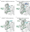
Fig. 4. Tail or 2nd head association has no effect on the ADP-bound conformation of the MT-associated Kif5b motor domain.
a ADP-bound motor domains of the Kif5bMoNeIAK and Kif5bMoNeXXX (semi-transparent) MT-associated models were superimposed (left). RMSDs were calculated and displayed on the Kif5bMoNeIAK model (right), with the Kif5bMoNeIAK tail shown in magenta. b MT-associated ADP-bound motor domains of the Kif5bMoNeIAK one (semi-transparent) and two-headed models were superimposed (left). RMSDs were calculated and displayed on the Kif5bMoNeIAK two-headed model (right) with the Kif5bMoNeIAK tail shown in magenta. c Overview of the nucleotide-pocket and switch-motif containing loops 9 and 11 (L9 and L11) in the ADP-bound Kif5bMoNeIAK one-headed model, with side chains for key mechanistic residues shown. Cryo-EM density is shown as semi-transparent gray and was filtered using DeepEMhancer. d, e Superimposition of the ADP-bound MT-associated motor domains of Kif5bMoNeXXX (semi-transparent) and d the Kif5bMoNeIAK one-headed model, e the Kif5bMoNeIAK two-headed model. A view of the nucleotide-binding site is shown, alongside key side chains involved in Mg2+ and ADP release. f, g Views of the 2nd (not MT-associated) head in the Kif5bMoNeIAK two-headed model in the presence of ADP. Cryo-EM density (semi-transparent) was low-pass filtered to 6 Å resolution for resolution-appropriate visualization. The position where density for an extended helix α4 and ordered L11 could be expected (but is absent here) is indicated in semi-transparent colors with dashed black outlines.
Fig. 5
Fig. 5. Tail or 2nd head association has no effect on the ATP-like conformation of the MT-associated Kif5b motor domain.
a Motor domains of the Kif5bMoNeIAK AMPPNP and MT-bound and ADP-AlF4 and tubulin-bound (semi-transparent, PDB code 4hna) models were superimposed (left). RMSDs were calculated and displayed on the Kif5bMoNeIAK model (right), with the Kif5bMoNeIAK tail shown in magenta. b MT-associated AMPPNP-bound motor domains of the Kif5bMoNeIAK one (semi-transparent) and two-headed models were superimposed (left). RMSDs were calculated and displayed on the Kif5bMoNeIAK two-headed model (right) with the Kif5bMoNeIAK tail shown in magenta and the NL-CC region shown in green. c Overview of the nucleotide-pocket and switch-motif containing loops 9 and 11 (L9 and L11) in the AMPPNP-bound Kif5bMoNeIAK one-headed model, with side chains for key conserved residues shown. Cryo-EM density is shown as semi-transparent gray and was filtered with DeepEMhancer. d, e Close-up view of residues involved in ATP hydrolysis, comparing d our MT-associated AMPPNP-bound Kif5bMoNeIAK motor domain (left) and e Eg5 motor domain (PDB code 3hqd). Both models are fitted into density (local resolution sharpened in Relion) for the MT-associated motor domain of AMPPNP-bound Kif5bMoNeIAK (mesh). The high resolution in the Eg5 motor domain crystal structure allows display of the two water molecules and associated bonding network (black dashed lines) that are involved in nucleophilic attack on the γ-phosphate. In the right-hand panel, Eg5 residue numbering is shown in black and Kif5b residue numbering in gray. f, g Views of the 2nd (not MT-associated) head in the Kif5bMoNeIAK two-headed model in the presence of AMPPNP. Cryo-EM density (semi-transparent) was low-pass filtered to 6 Å resolution for resolution-appropriate visualization. The position where density for an extended helix α4 and ordered L11 could be expected (but is absent here) is indicated in semi-transparent colors with dashed black outlines.
Fig. 6
Fig. 6. MT-associated Kif5bMoNeIAK undocks its neck linker in the presence of ADP.
ad Views of the neck linker and coiled-coil of the MT-associated head of Kif5bMoNeIAK bound to a, b AMPPNP or c, d ADP in (a, c) one- or (b, d) two-headed states, with corresponding density in semi-transparent gray. In (c) and (d), the position where density for a docked neck linker could be expected (but is absent here) is indicated in semi-transparent cyan. All cryo-EM densities shown in (ac) were filtered using DeepEMhancer. In (d), the displayed density for the 1st head was filtered using DeepEMhancer, while the density for the 2nd head displayed density was low-pass filtered to 6 Å resolution for resolution-appropriate visualization.
Fig. 7
Fig. 7. Model for a two-step structural mechanism of MT-stimulated ADP release.
Schematic illustrating a proposed two-step structural mechanism of MT-stimulated ADP release, showing movements of key SSEs and side chains relative to the static helix α2a and P-loop. MT binding triggers transition to an apo-like state, including helix α4 extension and twisting of core β-sheet (β-core) along with movements of overlying helix α3 and loop 9 elements. These movements dismantle a hydrogen bonding network involving D231 and T92 that coordinates Mg2+ through a water ‘cap’ (blue dashed lines). This drives Mg2+ out of the complex, with D231 forming a new bonding network with R190 and K91 (red dashed lines). These changes disrupt key bonds stabilizing ADP’s β-phosphate (cyan dashed lines), leaving ADP only weakly coordinated by remaining contacts with helix α2a and the P-loop, promoting its subsequent release mediated by loop 9 dynamics. α and β are α-phosphate and β-phosphate, respectively.

Similar articles

Cited by

References

    1. Hirokawa, N., Noda, Y., Tanaka, Y. & Niwa, S. Kinesin superfamily motor proteins and intracellular transport. Nat. Rev. Mol. Cell Biol.10, 682–696 (2009). - PubMed
    1. Mandal, A. & Drerup, C. M. Axonal transport and mitochondrial function in neurons. Front. Cell Neurosci.13, 373 (2019). - PMC - PubMed
    1. Schwarz, T. L. Mitochondrial trafficking in neurons. Cold Spring Harb. Perspect. Biol.10.1101/cshperspect.a011304 (2013). - PMC - PubMed
    1. Guedes-Dias, P. & Holzbaur, E. L. F. Axonal transport: driving synaptic function. Science366, eaaw9997 (2019). - PMC - PubMed
    1. Hollenbeck, P. J. & Swanson, J. A. Radial extension of macrophage tubular lysosomes supported by kinesin. Nature346, 864–866 (1990). - PubMed

LinkOut - more resources